Université Paris-Saclay, CEA, CNRS, Baobab, Neurospin, Gif-sur-Yvette, France.
Commissariat à l'Energie Atomique et aux Energies Alternatives (CEA), Département des Sciences du Vivant (DSV), Institut d'Imagerie Biomédicale (I2BM), MIRCen, France.
Neuroimage Clin. 2020;26:102211. doi: 10.1016/j.nicl.2020.102211. Epub 2020 Feb 13.
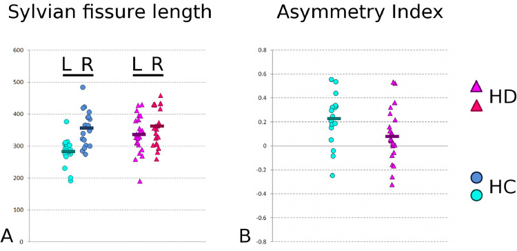
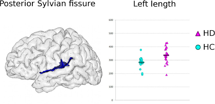
Huntington's disease (HD) is an inherited, autosomal dominant disorder that is characteristically thought of as a degenerative disorder. Despite cellular and molecular grounds suggesting HD could also impact normal development, there has been scarce systems-level data obtained from in vivo human studies supporting this hypothesis. Sulcus-specific morphometry analysis may help disentangle the contribution of coexisting neurodegenerative and neurodevelopmental processes, but such an approach has never been used in HD. Here, we investigated cortical sulcal depth, related to degenerative process, as well as cortical sulcal length, related to developmental process, in early-stage HD and age-matched healthy controls. This morphometric analysis revealed significant differences in the HD participants compared with the healthy controls bilaterally in the central and intra-parietal sulcus, but also in the left intermediate frontal sulcus and calcarine fissure. As the primary visual cortex is not connected to the striatum, the latter result adds to the increasing in vivo evidence for primary cortical degeneration in HD. Those sulcal measures that differed between HD and healthy populations were mainly atrophy-related, showing shallower sulci in HD. Conversely, the sulcal morphometry also revealed a crucial difference in the imprint of the Sylvian fissure that could not be related to loss of grey matter volume: an absence of asymmetry in the length of this fissure in HD. Strong asymmetry in that cortical region is typically observed in healthy development. As the formation of the Sylvian fissure appears early in utero, and marked asymmetry is specifically found in this area of the neocortex in newborns, this novel finding likely indicates the foetal timing of a disease-specific, genetic interplay with neurodevelopment.
亨廷顿病(HD)是一种遗传性、常染色体显性疾病,通常被认为是一种退行性疾病。尽管细胞和分子基础表明 HD 也可能影响正常发育,但从支持这一假说的体内人类研究中获得的系统水平数据却很少。沟回特异性形态测量分析可能有助于理清共存的神经退行性和神经发育过程的贡献,但这种方法从未在 HD 中使用过。在这里,我们研究了皮质沟回深度,与退行性过程有关,以及皮质沟回长度,与发育过程有关,在早期 HD 和年龄匹配的健康对照组中。这项形态测量分析显示,与健康对照组相比,HD 参与者双侧中央沟和顶内沟,以及左侧中间额沟和距状裂都有显著差异。由于初级视觉皮层与纹状体没有连接,因此后者的结果增加了越来越多的体内证据表明 HD 中存在原发性皮质退行性变。在 HD 和健康人群之间存在差异的那些脑回测量主要与萎缩有关,在 HD 中脑回较浅。相反,脑回形态测量还揭示了大脑外侧裂的一个关键差异,这不能与灰质体积的丧失联系起来:HD 中这条裂的长度没有不对称。在健康发育过程中,通常会观察到该皮质区域的强烈不对称。由于大脑外侧裂的形成发生在子宫内早期,并且在新生儿的新皮层中特定区域发现了明显的不对称,因此这一新发现可能表明存在一种疾病特异性的、与神经发育相关的遗传相互作用的胎儿时间。