Chen Yihong, Shi Ce, Zhou Lili, Huang Shenghai, Shen Meixiao, He Zhiyong
School of Ophthalmology and Optometry, Wenzhou Medical University, Wenzhou, China.
Department of Neurology, The Second Affiliated Hospital and Yuying Children's Hospital of Wenzhou Medical University, Wenzhou, China.
Front Neurol. 2020 Feb 11;11:35. doi: 10.3389/fneur.2020.00035. eCollection 2020.
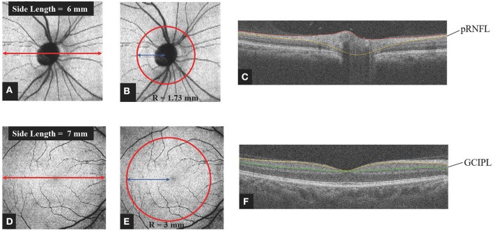
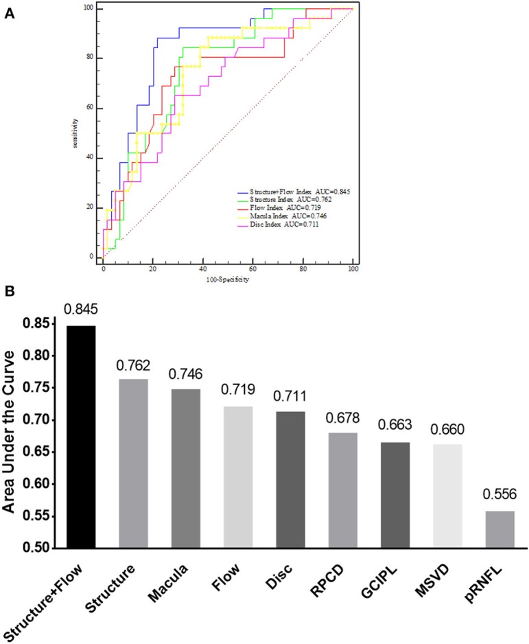
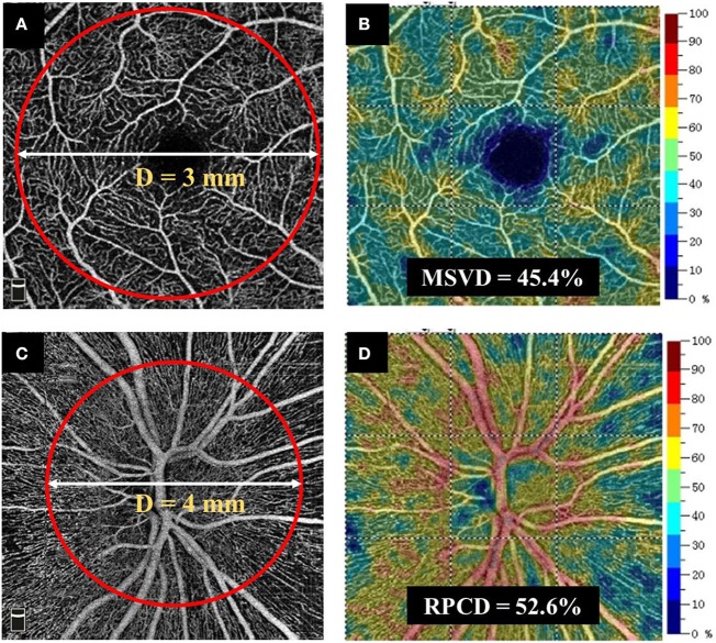
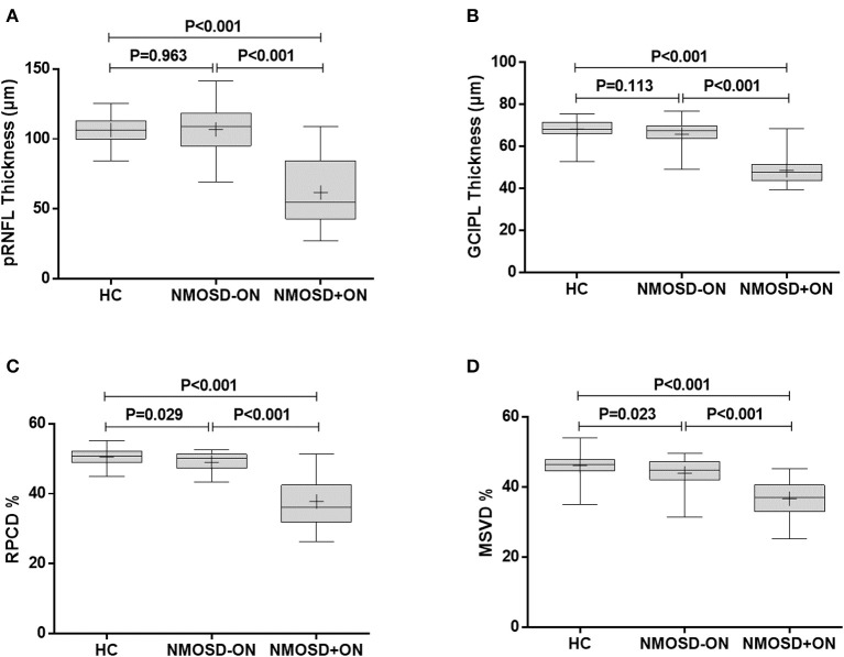
To use optical coherence tomography (OCT) and OCT angiography (OCT-A) to measure changes in the retinal structure and microvasculature of patients with aquaporin-4 antibody-positive, neuromyelitis optica spectrum disorder (NMOSD) with a history of optic neuritis (NMOSD+ON) and those without it (NMOSD-ON). A total of 27 aquaporin-4 antibody-positive NMOSD patients and 31 age- and gender-matched healthy control (HC) participants were included. In 27 NMOSD patients, 19 of them had a history of optic neuritis (ON) and 8 of them had no history of ON. Peripapillary retinal nerve fiber layer (pRNFL) thickness and macular ganglion cell and inner plexiform layer (GCIPL) thickness were measured by OCT. Radial peripapillary capillary density (RPCD) and macular superficial vessel density (MSVD) were measured by OCT-A. Comparisons of retinal structural and microvascular parameters between the cohorts were performed using generalized estimating equation (GEE) models. Diagnostic accuracy was evaluated by the area under the receiver operating characteristics curve (AROC). In NMOSD+ON eyes, the GCIPL and pRNFL thicknesses, 48.6 ± 7.1 and 61.7 ± 25.1 μm, respectively, were significantly thinner than in HC eyes ( < 0.001 for both). However, in NMOSD-ON eyes, the GCIPL and pRNFL thicknesses were not significantly thinner than in HC eyes ( > 0.05 for both). In NMOSD+ON eyes, the RPCD and MSVD, 37.8 ± 7.1 and 36.7 ± 5.0%, respectively, were significantly less dense than HC eyes ( < 0.001 for both). Similarly, the RPCD and MSVD in NMOSD-ON eyes, 49.0 ± 2.8 and 43.9 ± 4.2%, respectively, were also less dense than in HC eyes ( < 0.029 for RPCD, < 0.023 for MSVD). The highest AROC, 0.845 (sensitivity = 88.5%, specificity = 78.0%), was achieved by the logistic regression combination of all of the variables, i.e., pRNFL, GCIPL, RPCD, and MSVD. Retinal microvascular changes were present in NMOSD-ON eyes. The combination of retinal structural and microvascular parameters might be helpful to discriminate NMOSD-ON eyes from HC eyes.
使用光学相干断层扫描(OCT)和OCT血管造影(OCT-A)来测量水通道蛋白4抗体阳性、有视神经炎病史的视神经脊髓炎谱系障碍(NMOSD+ON)患者和无视神经炎病史的患者(NMOSD-ON)的视网膜结构和微血管变化。共纳入27例水通道蛋白4抗体阳性的NMOSD患者和31名年龄及性别匹配的健康对照(HC)参与者。在27例NMOSD患者中,19例有视神经炎(ON)病史,8例无ON病史。通过OCT测量视乳头周围视网膜神经纤维层(pRNFL)厚度以及黄斑神经节细胞和内丛状层(GCIPL)厚度。通过OCT-A测量视乳头周围径向毛细血管密度(RPCD)和黄斑浅表血管密度(MSVD)。使用广义估计方程(GEE)模型对各队列之间的视网膜结构和微血管参数进行比较。通过受试者操作特征曲线下面积(AROC)评估诊断准确性。在NMOSD+ON眼中,GCIPL和pRNFL厚度分别为48.6±7.1和61.7±25.1μm,显著薄于HC眼(两者均P<0.001)。然而,在NMOSD-ON眼中,GCIPL和pRNFL厚度并不显著薄于HC眼(两者均P>0.05)。在NMOSD+ON眼中,RPCD和MSVD分别为37.8±7.1和36.7±5.0%,密度显著低于HC眼(两者均P<0.001)。同样,NMOSD-ON眼中的RPCD和MSVD分别为49.0±2.8和43.9±4.2%,也低于HC眼(RPCD为P<0.029,MSVD为P<0.023)。所有变量(即pRNFL、GCIPL、RPCD和MSVD)的逻辑回归组合获得了最高的AROC,为0.845(敏感性=88.5%,特异性=78.0%)。视网膜微血管变化存在于NMOSD-ON眼中。视网膜结构和微血管参数的组合可能有助于区分NMOSD-ON眼和HC眼。