Bera Debabrata, Saggu Daljeet, Yalagudri Sachin, Kadel Jugal Kishor, Sarkar Rakesh, Devidutta Soumen, Christopher Johann, Pavri Behzad, Narasimhan Calambur
Dept of Electrophysiology, Care Hospitals, Hyderabad, India.
Dept of Rheumatology, Care Hospitals, Hyderabad, India.
Indian Pacing Electrophysiol J. 2020 May-Jun;20(3):83-90. doi: 10.1016/j.ipej.2020.02.003. Epub 2020 Feb 29.
Patients with outflow tract ventricular tachycardia (OTVT) with normal echocardiogram are labeled as idiopathic VT (IVT). However, a subset of these patients is subsequently diagnosed with underlying cardiac sarcoidosis (CS).
Whether electrocardiogram (ECG) abnormalities in sinus rhythm (SR) can differentiate underlying CS from IVT.
We retrospectively analyzed the SR-ECGs of 42 patients with OTVT/premature ventricular complexes (PVC) and normal echocardiography. All underwent advanced imaging with cardiac magnetic resonance (CMR)/FDG PET-CT for screening of CS. Twenty-two patients had significant abnormalities in cardiac imaging and subsequently had biopsy-proven CS (Cases). Twenty patients had normal imaging and were categorized as IVT (Controls). SR-ECGs of all patients were analyzed by 2 independent, blinded observers.
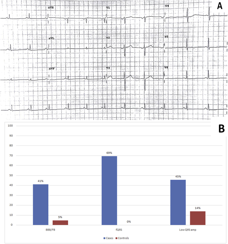
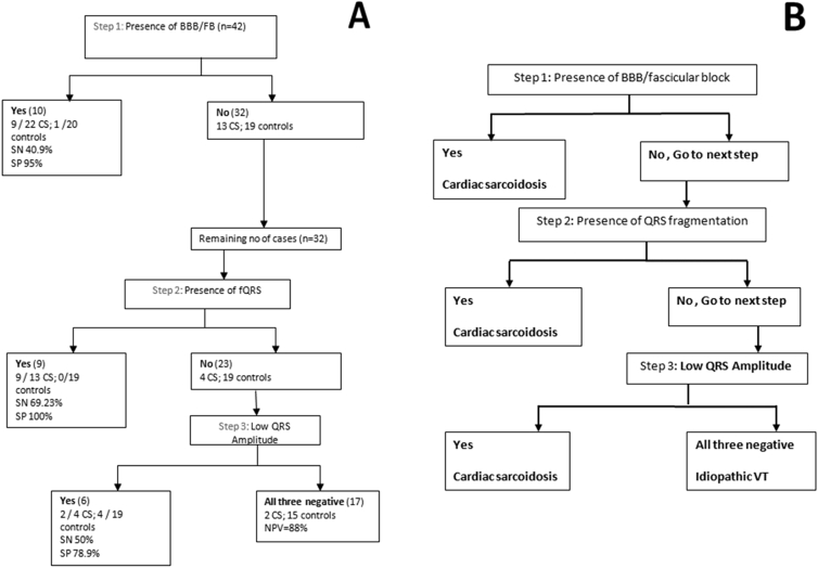
Baseline characteristics were comparable. Among the ECG features analyzed - fascicular (FB) or bundle branch block (BBB) was seen in 9/22 Cases vs. 1/20 controls (p = 0.01). Among patients without FB or BBB, fragmented QRS (fQRS) was present in 9/13 cases but in none of the controls (p < 0.001). Low voltage QRS was more often seen among cases as compared to controls (10/22 vs. 3/20 p = 0.03). A stepwise algorithm based on these 3 sets of ECG findings helped to diagnose CS among patients presenting with OTVT/PVC with sensitivity of 91%, specificity of 75%, a PPV of 80%, and a NPV of 88%.
In patients presenting with OTVT/PVC: FB/BBB, fQRS, and low QRS voltage on the baseline ECG were more often observed among patients with underlying CS as compared to true IVT. These findings may help to distinguish underlying CS among Cases presenting with OTVT/PVC.
超声心动图正常的流出道室性心动过速(OTVT)患者被归类为特发性室性心动过速(IVT)。然而,这些患者中有一部分随后被诊断为潜在的心脏结节病(CS)。
窦性心律(SR)时的心电图(ECG)异常是否能区分潜在的CS与IVT。
我们回顾性分析了42例OTVT/室性早搏(PVC)且超声心动图正常患者的SR-ECG。所有患者均接受心脏磁共振(CMR)/FDG PET-CT高级成像以筛查CS。22例患者心脏成像有显著异常,随后经活检证实为CS(病例组)。20例患者成像正常,归类为IVT(对照组)。所有患者的SR-ECG由2名独立、不知情的观察者进行分析。
基线特征具有可比性。在分析的ECG特征中,9/22例病例出现分支(FB)或束支传导阻滞(BBB),而对照组为1/20(p = 0.01)。在无FB或BBB的患者中,9/13例病例存在碎裂QRS波(fQRS),而对照组均无(p < 0.001)。与对照组相比,病例组中低电压QRS波更常见(10/22 vs. 3/20,p = 0.03)。基于这三组ECG结果的逐步算法有助于诊断OTVT/PVC患者中的CS,敏感性为91%,特异性为75%,阳性预测值为80%,阴性预测值为88%。
在OTVT/PVC患者中:与真正的IVT相比,潜在CS患者在基线ECG上更常出现FB/BBB、fQRS和低QRS电压。这些发现可能有助于在OTVT/PVC病例中区分潜在的CS。