Truttmann Anita C, Ginet Vanessa, Puyal Julien
Clinic of Neonatology, Department of Women, Mother and Child, University Hospital Center of Vaud, Lausanne, Switzerland.
Department of Fundamental Neurosciences, University of Lausanne, Lausanne, Switzerland.
Front Cell Dev Biol. 2020 Feb 18;8:27. doi: 10.3389/fcell.2020.00027. eCollection 2020.
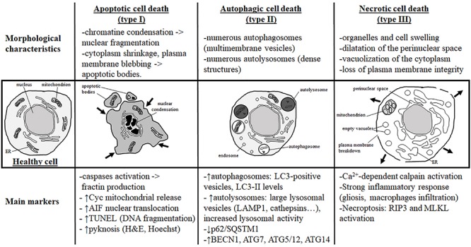
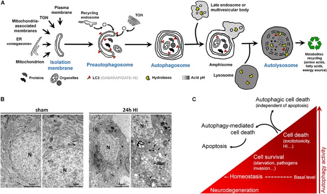
Despite tremendous advances in neonatal intensive care over the past 20 years, prematurity carries a high burden of neurological morbidity lasting lifelong. The term encephalopathy of prematurity (EoP) coined by Volpe in 2009 encompasses all aspects of the now known effects of prematurity on the immature brain, including altered and disturbed development as well as specific lesional hallmarks. Understanding the way cells are damaged is crucial to design brain protective strategies, and in this purpose, preclinical models largely contribute to improve the comprehension of the cell death mechanisms. While neuronal cell death has been deeply investigated and characterized in (hypoxic-ischemic) encephalopathy of the newborn at term, little is known about the types of cell death occurring in preterm brain injury. Three main different morphological cell death types are observed in the immature brain, specifically in models of hypoxic-ischemic encephalopathy, namely, necrotic, apoptotic, and autophagic cell death. Features of all three types may be present in the same dying neuron. In preterm brain injury, description of cell death types is sparse, and cell loss primarily concerns immature oligodendrocytes and, infrequently, neurons. In the present review, we first shortly discuss the different main severe preterm brain injury conditions that have been reported to involve cell death, including periventricular leucomalacia (PVL), diffuse white matter injury (dWMI), and intraventricular hemorrhages, as well as potentially harmful iatrogenic conditions linked to premature birth (anesthesia and caffeine therapy). Then, we present an overview of current evidence concerning cell death in both clinical human tissue data and preclinical models by focusing on studies investigating the presence of cell death allowing discriminating between the types of cell death involved. We conclude that, to improve brain protective strategies, not only apoptosis but also other cell death (such as regulated necrotic and autophagic) pathways now need to be investigated together in order to consider all cell death mechanisms involved in the pathogenesis of preterm brain damage.
尽管在过去20年新生儿重症监护方面取得了巨大进展,但早产带来的神经疾病负担依然沉重,会伴随终身。2009年沃尔普提出的早产儿脑病(EoP)一词涵盖了目前已知的早产对未成熟大脑影响的所有方面,包括发育改变和紊乱以及特定的损伤特征。了解细胞受损的方式对于设计脑保护策略至关重要,为此,临床前模型在很大程度上有助于增进对细胞死亡机制的理解。虽然在足月儿新生儿(缺氧缺血性)脑病中对神经元细胞死亡进行了深入研究和特征描述,但对于早产脑损伤中发生的细胞死亡类型却知之甚少。在未成熟大脑中,特别是在缺氧缺血性脑病模型中,观察到三种主要不同形态的细胞死亡类型,即坏死性、凋亡性和自噬性细胞死亡。所有这三种类型的特征可能存在于同一个濒死神经元中。在早产脑损伤中,细胞死亡类型的描述很少,细胞损失主要涉及未成熟少突胶质细胞,神经元则较少受累。在本综述中,我们首先简要讨论已报道涉及细胞死亡的不同主要严重早产脑损伤情况,包括脑室周围白质软化(PVL)、弥漫性白质损伤(dWMI)和脑室内出血,以及与早产相关的潜在有害医源性情况(麻醉和咖啡因治疗)。然后,我们通过关注研究细胞死亡存在情况以区分所涉及细胞死亡类型的研究,概述临床人体组织数据和临床前模型中有关细胞死亡的当前证据。我们得出结论,为了改进脑保护策略,现在不仅需要研究凋亡,还需要共同研究其他细胞死亡(如调节性坏死和自噬)途径,以便考虑早产脑损伤发病机制中涉及的所有细胞死亡机制。