Pertiwi Kartika R, de Boer Onno J, Gabriels Pauline A M, van der Wal Allard C
Department of Pathology, Amsterdam UMC, University of Amsterdam, Meibergdreef 9, 1105 AZ Amsterdam, Netherlands.
Department of Biology Education, Faculty of Mathematics and Natural Science, Yogyakarta State University, Jl. Colombo No. 1, Karang Malang, Caturtunggal, Kec. Depok, Kabupaten Sleman, Daerah Istimewa Yogyakarta 55281, Indonesia.
Int J Cardiol Heart Vasc. 2019 Nov 25;26:100439. doi: 10.1016/j.ijcha.2019.100439. eCollection 2020 Feb.
Coronary thrombosis is a process with unpredictable clinical outcome. Changes of thrombus composition overtime influence tissue repair and stabilization. We investigated rates of cell deaths and cell proliferation at different time points after initiation of thrombosis.
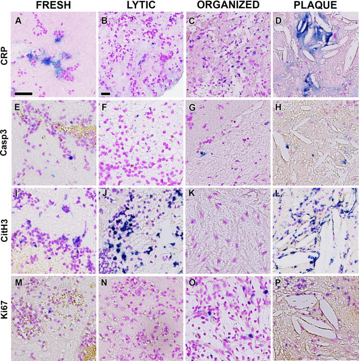
Thrombectomy aspirates of 55 myocardial infarction patients were selected and histomorphologically classified as fresh (25), lytic (25), partially fibrocellular (10), completely fibrocellular (10). Paraffin sections were immunostained with anti-(cleaved) caspase-3/Casp3 (apoptosis), Citrullinated histone/CitH 3 (etosis), C-reactive protein/CRP and Ki67 (proliferation) in combination with either Feulgen counterstaining (DNA) or cell markers for granulocytes, macrophages, SMCs, platelets and endothelium. Rates of apoptosis, etosis and proliferation were measured as a percentage of total number of immunopositive pixels versus total number of DNA positive pixels, while co-localization with cell markers was assessed by digital image analysis.
Positive staining of CitH3 was observed more frequently (93%) than Casp3 (70%), Ki67 (79%) or CRP (59%) (p < 0.05). Moreover, rate of etosis, found in granulocytes and macrophages, differed significantly among thrombi of different age, being higher in lytic (12.82) than in fresh (8.52) and late-organized (2.75) (p < 0.05). Such differences were not observed for the rates of apoptosis or cell proliferation related to thrombus age. CRP staining was present in fresh, lytic and organized thrombi, but did not reliably identify necrotic areas.
Different patterns of cell death and cell proliferation are noticed during progression of coronary thrombus overtime, but with significant differences for only etosis. Etosis could potentially serve as a biomarker for thrombus instability with clinical significance.
冠状动脉血栓形成是一个临床结局不可预测的过程。血栓成分随时间的变化会影响组织修复和稳定。我们研究了血栓形成开始后不同时间点的细胞死亡率和细胞增殖率。
选取55例心肌梗死患者的血栓切除术吸出物,进行组织形态学分类,分为新鲜血栓(25例)、溶解血栓(25例)、部分纤维细胞性血栓(10例)、完全纤维细胞性血栓(10例)。石蜡切片用抗(裂解)半胱天冬酶-3/Casp3(凋亡)、瓜氨酸化组蛋白/CitH 3(细胞程序性坏死)、C反应蛋白/CRP和Ki67(增殖)进行免疫染色,并结合福尔根复染(DNA)或粒细胞、巨噬细胞、平滑肌细胞、血小板和内皮细胞的细胞标志物。凋亡、细胞程序性坏死和增殖率以免疫阳性像素总数占DNA阳性像素总数的百分比来衡量,而与细胞标志物的共定位则通过数字图像分析进行评估。
观察到CitH3的阳性染色比Casp3(70%)、Ki67(79%)或CRP(59%)更频繁(93%)(p<0.05)。此外,在粒细胞和巨噬细胞中发现的细胞程序性坏死率在不同年龄的血栓中差异显著,溶解血栓(12.82)中的细胞程序性坏死率高于新鲜血栓(8.52)和晚期机化血栓(2.75)(p<0.05)。与血栓年龄相关的凋亡率或细胞增殖率未观察到此类差异。CRP染色存在于新鲜、溶解和机化血栓中,但不能可靠地识别坏死区域。
在冠状动脉血栓形成随时间进展的过程中,观察到不同的细胞死亡和细胞增殖模式,但仅细胞程序性坏死存在显著差异。细胞程序性坏死可能作为具有临床意义的血栓不稳定的生物标志物。