Bakshi Sabyasachi
Department of General Surgery, BSMCH, Bankura, India.
, Hooghly, 712103, West Bengal, India.
Surg Case Rep. 2020 Mar 5;6(1):47. doi: 10.1186/s40792-020-00810-3.
Pampiniform plexus thrombosis is a very rare disease (only less than 25 published cases are available till date), and it is a diagnostic dilemma. The present case is an unusual condition of an elderly gentleman who was finally diagnosed as a case of spontaneous thrombosis of bilateral pampiniform plexus and was managed conservatively. Literature was reviewed to explore potential etiologies and therapeutic strategies.
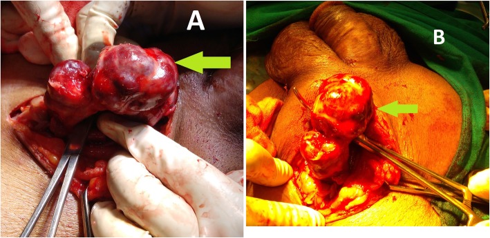
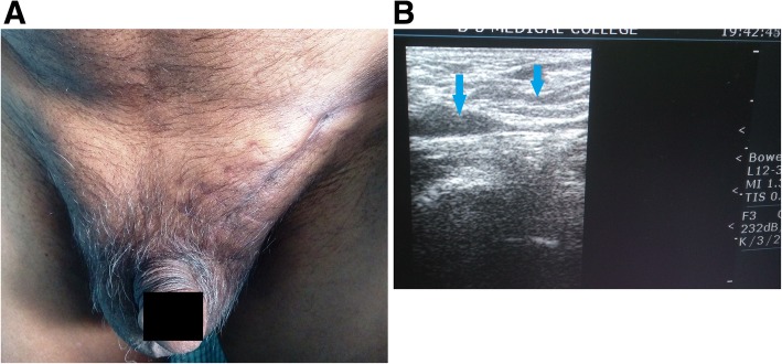
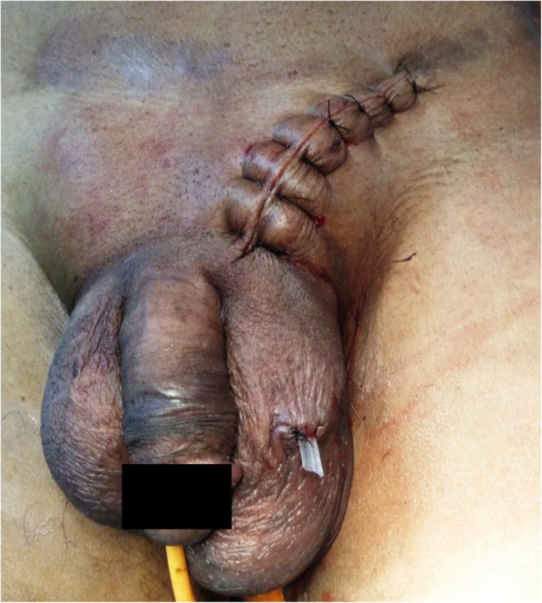
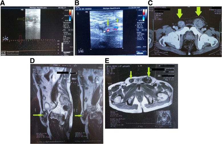
A 65-year-old afebrile gentleman, laborer (in brick industry), and non-smoker with no previous major health problems was admitted with swelling in the bilateral inguinal region. The swelling had started one and half months ago. He had developed severe pain over the swelling for last 1 day with tenderness and indurations. Neither he had history of previous surgeries, chronic cough, dysuria, prostatism, and trauma nor he presented any thrombogenic factors. There was no history of vomiting, abdominal pain, and obstipation. Physical examination revealed normotensive person with BMI of 22.5, was significant only for one tender, movable, and firm to hard 10 cm × 3 cm mass extending from the left deep inguinal ring up to the upper pole of the testis in the scrotum. Another 5 cm × 3 cm mass of similar characteristics was found extending from deep inguinal ring up to the root of the scrotum on right side. The testes and prostate were normal on palpation. On the contrary to preoperative USG, which clinched suspicion of incarcerated inguinal hernia, a thrombosed pampiniform plexus without any evidence of hernia sac was found on the left side during inguino-scrotal exploration. Wound was closed without doing any further procedure. Contralateral inguino-scrotal exploration was spared considering same nature of disease. Postoperative Doppler ultrasonography confirmed the diagnosis of bilateral thrombosed pampiniform plexus. MDCT of whole abdomen revealed no abnormality other than bilateral spermatic cord thrombosis. Blood thrombophilia screening came normal. The subject had an uneventful postoperative hospital course. With 2 years of follow-up, the gentleman is doing well, remaining asymptomatic and had returned to his usual life.
Due to extreme rarity, spontaneous thrombosis of the pampiniform plexus may be a diagnostic dilemma and requires a high index of suspicion. Doppler ultrasound is the initial investigation of choice. In the absence of other concomitant disease, beginning the treatment conservatively instead of excising the thrombosed segment is more suitable.
精索静脉曲张血栓形成是一种非常罕见的疾病(截至目前仅有不到25例已发表的病例),并且是一种诊断难题。本病例是一位老年男性的特殊情况,最终被诊断为双侧精索静脉曲张自发性血栓形成,并采用保守治疗。回顾文献以探讨潜在病因和治疗策略。
一名65岁的男性,从事体力劳动(砖厂工人),不吸烟,既往无重大健康问题,因双侧腹股沟区肿胀入院。肿胀始于一个半月前。在过去1天里,他在肿胀部位出现了严重疼痛,伴有压痛和硬结。他既无既往手术史、慢性咳嗽、排尿困难、前列腺疾病和外伤史,也未表现出任何血栓形成因素。无呕吐、腹痛和便秘史。体格检查显示血压正常,BMI为22.5,仅发现一个压痛、可移动、质地硬的10厘米×3厘米肿块,从左侧腹股沟深环延伸至阴囊内睾丸的上极。右侧发现另一个大小为5厘米×3厘米、具有相似特征的肿块,从腹股沟深环延伸至阴囊根部。触诊时睾丸和前列腺正常。与术前超声检查结果相反,术前超声检查怀疑为嵌顿性腹股沟疝,但在腹股沟阴囊探查时,左侧发现了一个血栓形成的精索静脉曲张,未发现任何疝囊迹象。伤口未进行进一步处理即予缝合。考虑到疾病性质相同,对侧腹股沟阴囊探查未进行。术后多普勒超声检查证实了双侧精索静脉曲张血栓形成的诊断。全腹MDCT检查除双侧精索静脉曲张血栓形成外未发现其他异常。血液血栓形成倾向筛查结果正常。该患者术后住院过程顺利。经过2年的随访,该男性情况良好,无症状,已恢复正常生活。
由于极为罕见,精索静脉曲张自发性血栓形成可能是一个诊断难题,需要高度怀疑。多普勒超声是首选的初步检查方法。在没有其他伴随疾病的情况下,保守治疗而非切除血栓形成的节段更为合适。