• 文献检索
  • 文档翻译
  • 深度研究
  • 学术资讯
  • Suppr Zotero 插件Zotero 插件
  • 邀请有礼
  • 套餐&价格
  • 历史记录
应用&插件
Suppr Zotero 插件Zotero 插件浏览器插件Mac 客户端Windows 客户端微信小程序
定价
高级版会员购买积分包购买API积分包
服务
文献检索文档翻译深度研究API 文档MCP 服务
关于我们
关于 Suppr公司介绍联系我们用户协议隐私条款
关注我们

Suppr 超能文献

核心技术专利:CN118964589B侵权必究
粤ICP备2023148730 号-1Suppr @ 2026

文献检索

告别复杂PubMed语法,用中文像聊天一样搜索,搜遍4000万医学文献。AI智能推荐,让科研检索更轻松。

立即免费搜索

文件翻译

保留排版,准确专业,支持PDF/Word/PPT等文件格式,支持 12+语言互译。

免费翻译文档

深度研究

AI帮你快速写综述,25分钟生成高质量综述,智能提取关键信息,辅助科研写作。

立即免费体验

血管免疫母细胞性T细胞淋巴瘤和霍奇金病中PD-L1阳性细胞的免疫表型分析。

Immunophenotyping of the PD-L1-positive cells in angioimmunoblastic T cell lymphoma and Hodgkin disease.

作者信息

Tiemann Markus, Samoilova Vera, Atiakshin Dmitri, Buchwalow Igor

机构信息

Institute for Hematopathology, Hamburg, Germany.

Research Institute of Experimental Biology and Medicine, Burdenko Voronezh State Medical University, Voronezh, Russia.

出版信息

BMC Res Notes. 2020 Mar 7;13(1):139. doi: 10.1186/s13104-020-04975-w.

DOI:10.1186/s13104-020-04975-w
PMID:32143684
原文链接:https://pmc.ncbi.nlm.nih.gov/articles/PMC7060537/
Abstract

OBJECTIVE

Programmed death-1 (PD-1) and its ligand PD-L1 are now used as predictive biomarkers to guide clinical decisions. Precise characterization of PD-L1-positive cells may contribute to our knowledge of which patients derive benefit from the PD-L1 blockade therapy.

RESULTS

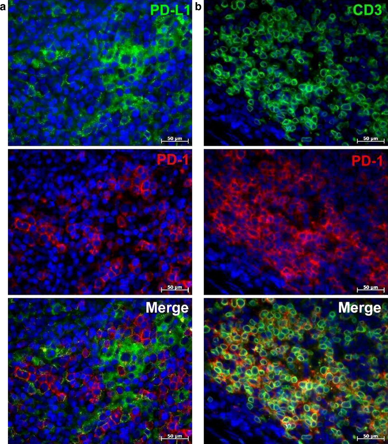
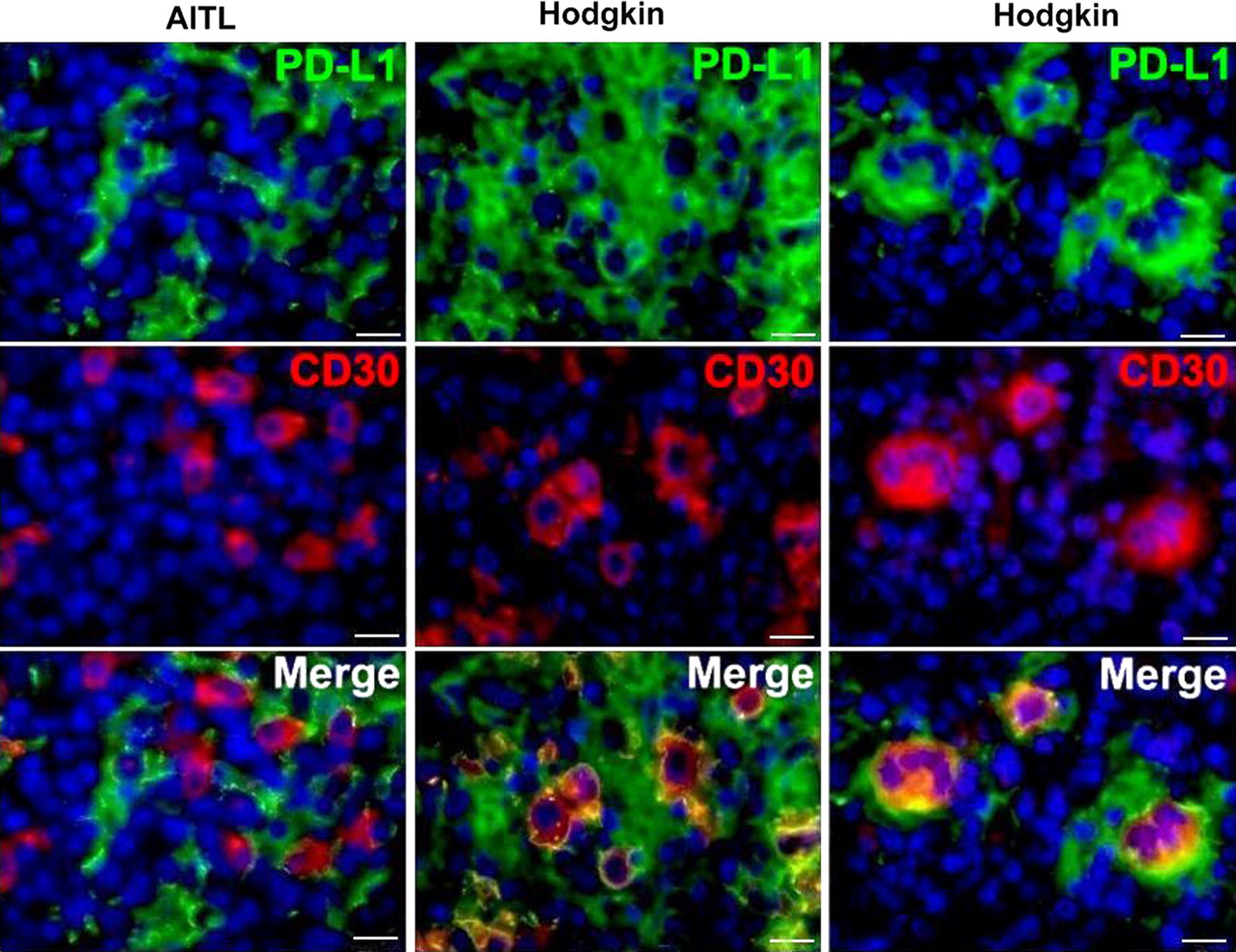
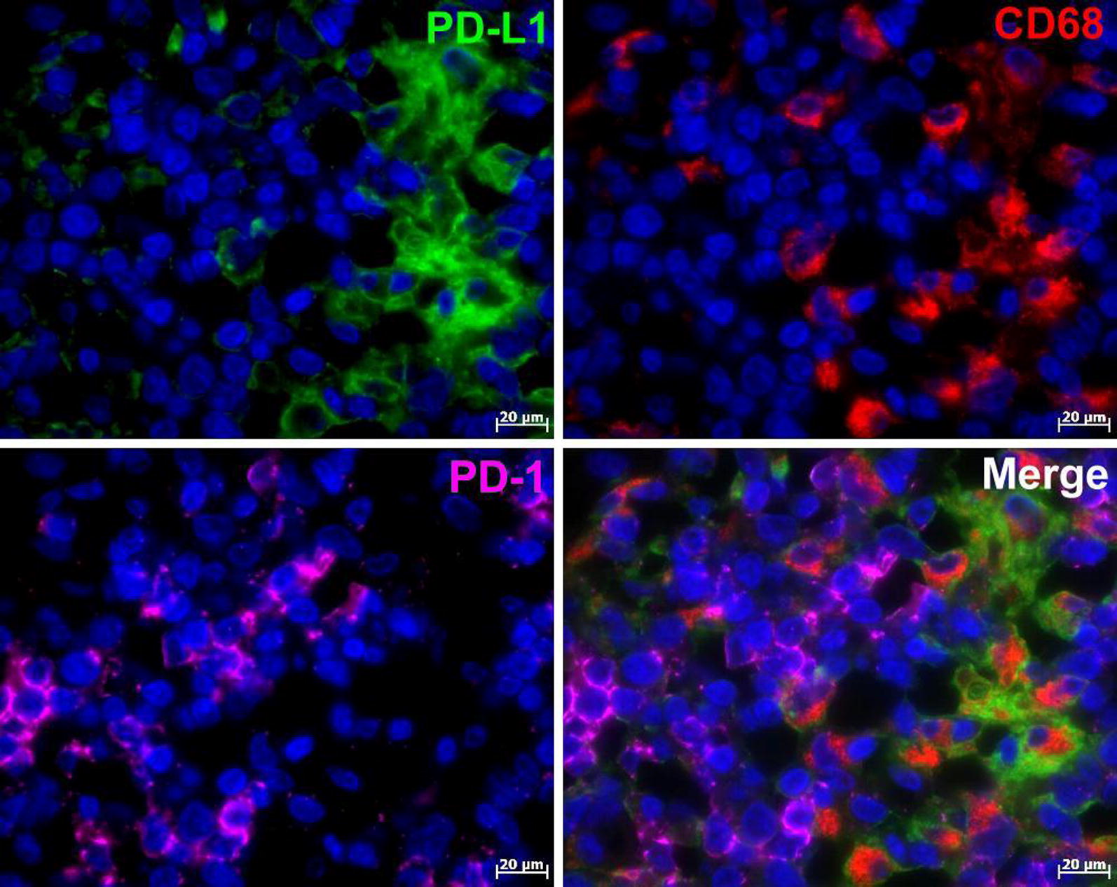
To address this issue, we performed immunophenotyping of PD-L1-positive cells in Hodgkin lymphoma and in angioimmunoblastic T cell lymphoma (AITL) employing multiple immunofluorescent immunolabeling. We found that PD-L1-positive cells and PD-1-positive cells both in Hodgkin lymphoma and in AITL belong to two completely different cell lineages. In both lymphomas, PD-1 was found exclusively in T-lymphocytes, whereas PD-L1 was revealed in the tumor microenvironment cells including macrophages. PD-L1 was also detected in CD30-positive cells in Hodgkin lymphoma but not in AITL. The marker of B-cell lineage, CD20, was not detectable in PD-L1-positive cells both in AITL and in Hodgkin. Our study highlights the importance of comprehensive assessment of PD-1/PD-L1 regulatory pathways for employing PD-L1 as a predictive biomarker in clinical practice. PD-L1-antibody therapy is proven in Hodgkin lymphoma. Comparative immunophenotyping of the PD-1/PD-L1 axis provides a support for attempts to prove this principle also for AITL.

摘要

目的

程序性死亡蛋白1(PD-1)及其配体PD-L1现被用作预测生物标志物以指导临床决策。对PD-L1阳性细胞进行精确表征可能有助于我们了解哪些患者能从PD-L1阻断治疗中获益。

结果

为解决这一问题,我们采用多重免疫荧光免疫标记技术对霍奇金淋巴瘤和血管免疫母细胞性T细胞淋巴瘤(AITL)中的PD-L1阳性细胞进行了免疫表型分析。我们发现,霍奇金淋巴瘤和AITL中的PD-L1阳性细胞和PD-1阳性细胞均属于两个完全不同的细胞谱系。在这两种淋巴瘤中,PD-1仅在T淋巴细胞中发现,而PD-L1则在包括巨噬细胞在内的肿瘤微环境细胞中显示。在霍奇金淋巴瘤的CD30阳性细胞中也检测到了PD-L1,但在AITL中未检测到。在AITL和霍奇金淋巴瘤的PD-L1阳性细胞中均未检测到B细胞谱系标志物CD20。我们的研究强调了全面评估PD-1/PD-L1调节通路对于在临床实践中采用PD-L1作为预测生物标志物的重要性。PD-L1抗体治疗在霍奇金淋巴瘤中已得到证实。对PD-1/PD-L1轴进行比较性免疫表型分析为尝试在AITL中也证明这一原理提供了支持。

https://cdn.ncbi.nlm.nih.gov/pmc/blobs/317a/7060537/8fb378554d5e/13104_2020_4975_Fig3_HTML.jpg
https://cdn.ncbi.nlm.nih.gov/pmc/blobs/317a/7060537/0acd8ed6f553/13104_2020_4975_Fig1_HTML.jpg
https://cdn.ncbi.nlm.nih.gov/pmc/blobs/317a/7060537/8ff4cd13ad43/13104_2020_4975_Fig2_HTML.jpg
https://cdn.ncbi.nlm.nih.gov/pmc/blobs/317a/7060537/8fb378554d5e/13104_2020_4975_Fig3_HTML.jpg
https://cdn.ncbi.nlm.nih.gov/pmc/blobs/317a/7060537/0acd8ed6f553/13104_2020_4975_Fig1_HTML.jpg
https://cdn.ncbi.nlm.nih.gov/pmc/blobs/317a/7060537/8ff4cd13ad43/13104_2020_4975_Fig2_HTML.jpg
https://cdn.ncbi.nlm.nih.gov/pmc/blobs/317a/7060537/8fb378554d5e/13104_2020_4975_Fig3_HTML.jpg

相似文献

1
Immunophenotyping of the PD-L1-positive cells in angioimmunoblastic T cell lymphoma and Hodgkin disease.血管免疫母细胞性T细胞淋巴瘤和霍奇金病中PD-L1阳性细胞的免疫表型分析。
BMC Res Notes. 2020 Mar 7;13(1):139. doi: 10.1186/s13104-020-04975-w.
2
Clinicopathological features of programmed cell death-1 and programmed cell death-ligand-1 expression in the tumor cells and tumor microenvironment of angioimmunoblastic T cell lymphoma and peripheral T cell lymphoma not otherwise specified.血管免疫母细胞性 T 细胞淋巴瘤和未特指外周 T 细胞淋巴瘤肿瘤细胞及肿瘤微环境中程序性细胞死亡受体-1 和程序性细胞死亡配体-1 表达的临床病理特征。
Virchows Arch. 2020 Jul;477(1):131-142. doi: 10.1007/s00428-020-02790-z. Epub 2020 Mar 13.
3
CD30 Expression by B and T Cells: A Frequent Finding in Angioimmunoblastic T-Cell Lymphoma and Peripheral T-Cell Lymphoma-Not Otherwise Specified.B细胞和T细胞的CD30表达:血管免疫母细胞性T细胞淋巴瘤和外周T细胞淋巴瘤(非特指型)中的常见现象
Am J Surg Pathol. 2016 Mar;40(3):378-85. doi: 10.1097/PAS.0000000000000571.
4
Major Histocompatibility Complex Class II and Programmed Death Ligand 1 Expression Predict Outcome After Programmed Death 1 Blockade in Classic Hodgkin Lymphoma.主要组织相容性复合体 II 类和程序性死亡配体 1 表达预测经典霍奇金淋巴瘤接受程序性死亡 1 阻断后的结局。
J Clin Oncol. 2018 Apr 1;36(10):942-950. doi: 10.1200/JCO.2017.77.3994. Epub 2018 Feb 2.
5
Profiling the Tumour Immune Microenvironment in Pancreatic Neuroendocrine Neoplasms with Multispectral Imaging Indicates Distinct Subpopulation Characteristics Concordant with WHO 2017 Classification.多光谱成像分析胰腺神经内分泌肿瘤的肿瘤免疫微环境,表明与 WHO 2017 分类一致的不同亚群特征。
Sci Rep. 2018 Sep 3;8(1):13166. doi: 10.1038/s41598-018-31383-9.
6
Enhanced PD-L1 expression on tumor cells in primary CD30-positive cutaneous large T-cell lymphoma: a report of lymph node lesions of four cases.原发性 CD30 阳性皮肤大 T 细胞淋巴瘤中肿瘤细胞 PD-L1 表达增强:4 例淋巴结病变报告。
J Clin Exp Hematop. 2023;63(1):49-57. doi: 10.3960/jslrt.22042.
7
PD-1-PD-L1 immune-checkpoint blockade in malignant lymphomas.恶性淋巴瘤中的PD-1-PD-L1免疫检查点阻断
Ann Hematol. 2018 Feb;97(2):229-237. doi: 10.1007/s00277-017-3176-6. Epub 2017 Nov 11.
8
Precursor cells and implications of a T-cell inflamed immune response in the pre-malignant setting in Hodgkin lymphoma.霍奇金淋巴瘤恶性前阶段的 T 细胞炎症免疫反应的前体细胞及其意义。
Immunobiology. 2020 Jan;225(1):151872. doi: 10.1016/j.imbio.2019.11.007. Epub 2019 Nov 25.
9
PD-1/PD-L1 Pathway and Its Blockade in Patients with Classic Hodgkin Lymphoma and Non-Hodgkin Large-Cell Lymphomas.PD-1/PD-L1 通路及其阻断在经典霍奇金淋巴瘤和非霍奇金大细胞淋巴瘤患者中的作用。
Curr Hematol Malig Rep. 2020 Aug;15(4):372-381. doi: 10.1007/s11899-020-00589-y.
10
MiR155 sensitized B-lymphoma cells to anti-PD-L1 antibody via PD-1/PD-L1-mediated lymphoma cell interaction with CD8+T cells.miR155 通过 PD-1/PD-L1 介导的淋巴瘤细胞与 CD8+T 细胞相互作用使 B 细胞淋巴瘤细胞对抗 PD-L1 抗体敏感。
Mol Cancer. 2019 Mar 30;18(1):54. doi: 10.1186/s12943-019-0977-3.

引用本文的文献

1
New insights into epigenetic regulation of resistance to PD-1/PD-L1 blockade cancer immunotherapy: mechanisms and therapeutic opportunities.对PD-1/PD-L1阻断癌症免疫疗法耐药性的表观遗传调控的新见解:机制与治疗机会
Exp Hematol Oncol. 2022 Nov 16;11(1):101. doi: 10.1186/s40164-022-00356-0.
2
Expression of immune checkpoint molecules programmed death protein 1, programmed death-ligand 1 and inducible T-cell co-stimulator in mycosis fungoides and Sézary syndrome: association with disease stage and clinical outcome.蕈样肉芽肿和塞扎里综合征中免疫检查点分子程序性死亡蛋白 1、程序性死亡配体 1 和诱导型 T 细胞共刺激分子的表达:与疾病阶段和临床结局的关联。
Br J Dermatol. 2022 Aug;187(2):234-243. doi: 10.1111/bjd.21063. Epub 2022 May 10.
3

本文引用的文献

1
[Predictive value of PD-L1 diagnostics].[程序性死亡受体配体1(PD-L1)诊断的预测价值]
Pathologe. 2018 Nov;39(6):498-519. doi: 10.1007/s00292-018-0507-x.
2
Biomarkers of response to immune checkpoint blockade in cancer treatment.癌症治疗中免疫检查点阻断反应的生物标志物。
Crit Rev Oncol Hematol. 2018 Oct;130:108-120. doi: 10.1016/j.critrevonc.2018.07.010. Epub 2018 Aug 3.
3
Multiple immunolabeling with antibodies from the same host species in combination with tyramide signal amplification.使用来自同一宿主物种的抗体并结合酪胺信号放大进行多重免疫标记。
Large cell morphology, CMYC+ tumour cells, and PD-1+ tumour cell/intense PD-L1+ cell reactions are important prognostic factors in nodal peripheral T-cell lymphomas with T follicular helper markers.
大细胞形态、CMYC+肿瘤细胞和 PD-1+肿瘤细胞/强烈 PD-L1+细胞反应是具有滤泡辅助 T 细胞标志物的结外 T 细胞淋巴瘤的重要预后因素。
Diagn Pathol. 2021 Nov 6;16(1):101. doi: 10.1186/s13000-021-01163-7.
Acta Histochem. 2018 Jul;120(5):405-411. doi: 10.1016/j.acthis.2018.05.002. Epub 2018 May 5.
4
PD-1 expression on tumor-specific T cells: Friend or foe for immunotherapy?肿瘤特异性T细胞上的PD-1表达:免疫治疗的友还是敌?
Oncoimmunology. 2017 Sep 14;7(1):e1364828. doi: 10.1080/2162402X.2017.1364828. eCollection 2017.
5
Signaling pathways and immune evasion mechanisms in classical Hodgkin lymphoma.经典型霍奇金淋巴瘤中的信号通路和免疫逃逸机制。
Hematology Am Soc Hematol Educ Program. 2017 Dec 8;2017(1):310-316. doi: 10.1182/asheducation-2017.1.310.
6
Identification of autofluorescent cells in human angioimmunoblastic T-cell lymphoma.人血管免疫母细胞性T细胞淋巴瘤中自发荧光细胞的鉴定
Histochem Cell Biol. 2018 Feb;149(2):169-177. doi: 10.1007/s00418-017-1624-y. Epub 2017 Dec 2.
7
Signaling pathways and immune evasion mechanisms in classical Hodgkin lymphoma.经典型霍奇金淋巴瘤中的信号通路与免疫逃逸机制
Blood. 2017 Nov 23;130(21):2265-2270. doi: 10.1182/blood-2017-06-781989.
8
Programmed death-1 ligands PD-L1 and PD-L2 show distinctive and restricted patterns of expression in lymphoma subtypes.程序性死亡受体-1 配体 PD-L1 和 PD-L2 在淋巴瘤亚型中表现出独特和受限的表达模式。
Hum Pathol. 2018 Jan;71:91-99. doi: 10.1016/j.humpath.2017.10.029. Epub 2017 Nov 6.
9
PD-1 expression and clinical PD-1 blockade in B-cell lymphomas.PD-1 表达与 B 细胞淋巴瘤的临床 PD-1 阻断
Blood. 2018 Jan 4;131(1):68-83. doi: 10.1182/blood-2017-07-740993. Epub 2017 Nov 8.
10
PD-1 and PD-L1 Checkpoint Signaling Inhibition for Cancer Immunotherapy: Mechanism, Combinations, and Clinical Outcome.用于癌症免疫治疗的PD-1和PD-L1检查点信号抑制:机制、联合应用及临床结果
Front Pharmacol. 2017 Aug 23;8:561. doi: 10.3389/fphar.2017.00561. eCollection 2017.