Yekula Anudeep, Gupta Mihir, Coley Nicholas, U Hoi Sang
Department of Neurosurgery, Massachusetts General Hospital Harvard Medical School, 185 Cambridge Street, Richard B. Simches Building 3rd Floor, Boston, MA, 02114, United States.
Departments of Neurosurgery, UCSD Health, 9300 Campus Point Drive #MC7893, La Jolla, CA, 92037-1300, United States.
Int J Surg Case Rep. 2020;68:124-128. doi: 10.1016/j.ijscr.2020.02.046. Epub 2020 Feb 28.
H3K27M-mutant diffuse midline glioma is a recently classified unique entity predominantly affecting pediatric patients and rarely adults. The clinicopathologic features in adults remain poorly characterized.
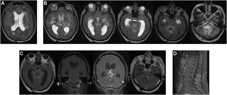
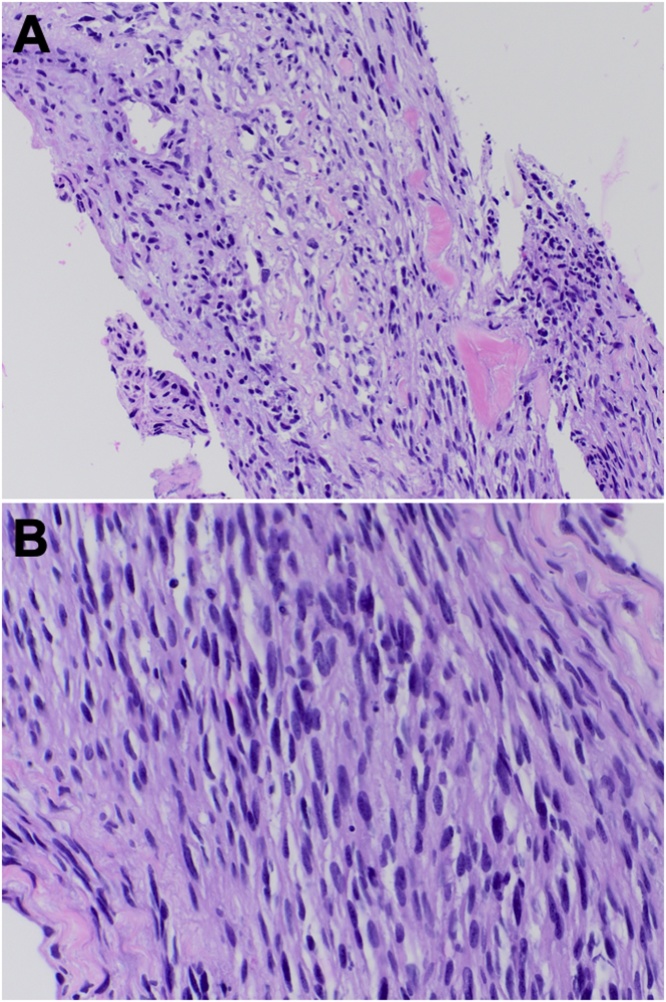
A 36-year-old man presented with subacute progressive cognitive and visual deterioration, and hydrocephalus requiring ventricular shunting. MRI revealed a diffusely infiltrating lesion with a gliomatosis cerebri growth pattern, multiple foci of contrast enhancement, and diffuse leptomeningeal involvement. Suboccipital craniotomy with exploration of the posterior fossa revealed a subtle capsular lesion infiltrating into the choroid plexus. Although histologically low-grade, the tumor was found to have an H3K27 M mutation establishing the diagnosis.
In spite of diverse clinicopathologic characteristics, H3K27M-mutant diffuse midline gliomas are incurable, WHO grade IV lesions with poor prognosis. We discuss our case in the context of a review of published reports of H3K27-mutant diffuse midline gliomas in adults. Findings late in the disease course may mimic inflammatory or infectious pathologies radiographically, and low-grade infiltrative neoplasms histologically.
The diverse clinical, radiographic and molecular features of H3K27M-mutant diffuse midline gliomas in adults remain to be completely characterized. A high index of suspicion is required to avoid missing the diagnosis. Early biopsy and detailed molecular characterization are critical for accurate diagnosis and patient counseling.
H3K27M突变型弥漫性中线胶质瘤是一种最近分类的独特实体,主要影响儿童患者,成人罕见。成人的临床病理特征仍不清楚。
一名36岁男性,表现为亚急性进行性认知和视力减退,以及需要脑室分流的脑积水。MRI显示一个弥漫性浸润性病变,呈大脑胶质瘤病生长模式,多个强化灶,以及弥漫性软脑膜受累。枕下开颅并探查后颅窝发现一个细微的包膜病变浸润至脉络丛。尽管组织学上为低级别,但肿瘤被发现有H3K27M突变,从而确立诊断。
尽管有不同的临床病理特征,H3K27M突变型弥漫性中线胶质瘤是无法治愈的WHO IV级病变,预后不良。我们在回顾已发表的成人H3K27突变型弥漫性中线胶质瘤报告的背景下讨论我们的病例。病程晚期的表现可能在影像学上类似炎症或感染性病变,在组织学上类似低级别浸润性肿瘤。
成人H3K27M突变型弥漫性中线胶质瘤多样的临床、影像学和分子特征仍有待完全明确。需要高度怀疑指数以避免漏诊。早期活检和详细的分子特征分析对于准确诊断和患者咨询至关重要。