Ucak Turgay, Unver Ethem
Department of Ophthalmology, Faculty of Medicine, Erzincan University, Erzincan, Turkey.
Department of Pulmonology, Faculty of Medicine, Erzincan University, Erzincan, Turkey.
J Ophthalmol. 2020 Jan 10;2020:4034382. doi: 10.1155/2020/4034382. eCollection 2020.
To analyze the effects of obstructive sleep apnea syndrome (OSAS) on ocular parameters and determine the alterations in macular vasculature by optical coherence tomography-angiography (OCT-A) in patients with different stages of OSAS.
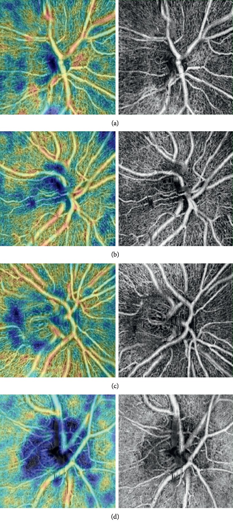
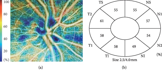
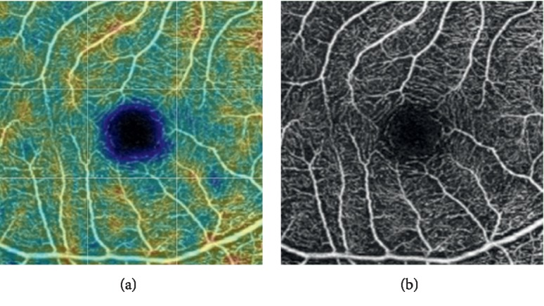
All the participants underwent a full ophthalmological examination. Using the macular OCT-A scans, the retinal peripapillary capillary plexus (RPCP), foveal avascular zone (FAZ), and superficial and deep vessel densities were recorded.
A total of 77 patients (154 eyes) with OSAS and 27 control cases (54 eyes) were included in this prospective study. Of the OSAS patients, 27 had mild, 24 had moderate, and 26 had severe disease. The intraocular pressure (IOP) values were significantly higher in the severe OSAS group than the control cases ( = 0.001). The average retinal nerve fiber layer (RNFL) thickness and the RNFL thickness of the temporal and inferior quadrants were significantly lower in the severe OSAS group compared with the control cases ( = 0.001). The average retinal nerve fiber layer (RNFL) thickness and the RNFL thickness of the temporal and inferior quadrants were significantly lower in the severe OSAS group compared with the control cases ( = 0.001). The average retinal nerve fiber layer (RNFL) thickness and the RNFL thickness of the temporal and inferior quadrants were significantly lower in the severe OSAS group compared with the control cases (.
Decreased vascular structures and increased FAZ may also be associated with the disease severity in OSAS and may be the main pathophysiological mechanisms in ocular alterations, which should be investigated in further studies.
分析阻塞性睡眠呼吸暂停综合征(OSAS)对眼部参数的影响,并通过光学相干断层扫描血管造影(OCT-A)确定不同阶段OSAS患者黄斑区血管系统的变化。
所有参与者均接受了全面的眼科检查。使用黄斑OCT-A扫描记录视网膜乳头周围毛细血管丛(RPCP)、黄斑无血管区(FAZ)以及浅表和深部血管密度。
本前瞻性研究纳入了77例(154只眼)OSAS患者和27例对照病例(54只眼)。在OSAS患者中,27例为轻度,24例为中度,26例为重度。重度OSAS组的眼压(IOP)值显著高于对照病例(P = 0.001)。与对照病例相比,重度OSAS组的平均视网膜神经纤维层(RNFL)厚度以及颞侧和下方象限的RNFL厚度显著降低(P = 0.001)。与对照病例相比,重度OSAS组的平均视网膜神经纤维层(RNFL)厚度以及颞侧和下方象限的RNFL厚度显著降低(P = 0.001)。与对照病例相比,重度OSAS组的平均视网膜神经纤维层(RNFL)厚度以及颞侧和下方象限的RNFL厚度显著降低(P <.
血管结构减少和FAZ增加也可能与OSAS的疾病严重程度相关,并且可能是眼部改变的主要病理生理机制,应在进一步研究中进行探讨。