• 文献检索
  • 文档翻译
  • 深度研究
  • 学术资讯
  • Suppr Zotero 插件Zotero 插件
  • 邀请有礼
  • 套餐&价格
  • 历史记录
应用&插件
Suppr Zotero 插件Zotero 插件浏览器插件Mac 客户端Windows 客户端微信小程序
定价
高级版会员购买积分包购买API积分包
服务
文献检索文档翻译深度研究API 文档MCP 服务
关于我们
关于 Suppr公司介绍联系我们用户协议隐私条款
关注我们

Suppr 超能文献

核心技术专利:CN118964589B侵权必究
粤ICP备2023148730 号-1Suppr @ 2026

文献检索

告别复杂PubMed语法,用中文像聊天一样搜索,搜遍4000万医学文献。AI智能推荐,让科研检索更轻松。

立即免费搜索

文件翻译

保留排版,准确专业,支持PDF/Word/PPT等文件格式,支持 12+语言互译。

免费翻译文档

深度研究

AI帮你快速写综述,25分钟生成高质量综述,智能提取关键信息,辅助科研写作。

立即免费体验

下颌舌骨嵴的维度和位置:锥形束 CT 对性别和骨骼形态的比较。

Dimension and Location of the Mandibular Lingula: Comparisons of Gender and Skeletal Patterns Using Cone-Beam Computed Tomography.

机构信息

Graduate Institute of Dental Sciences, School of Dental Medicine, Kaohsiung Medical University, Kaohsiung, Taiwan.

Department of Dentistry, Kaohsiung Municipal Ta-Tung Hospital, Kaohsiung, Taiwan.

出版信息

Biomed Res Int. 2020 Feb 13;2020:2571534. doi: 10.1155/2020/2571534. eCollection 2020.

DOI:10.1155/2020/2571534
PMID:32149093
原文链接:https://pmc.ncbi.nlm.nih.gov/articles/PMC7042528/
Abstract

OBJECTIVE

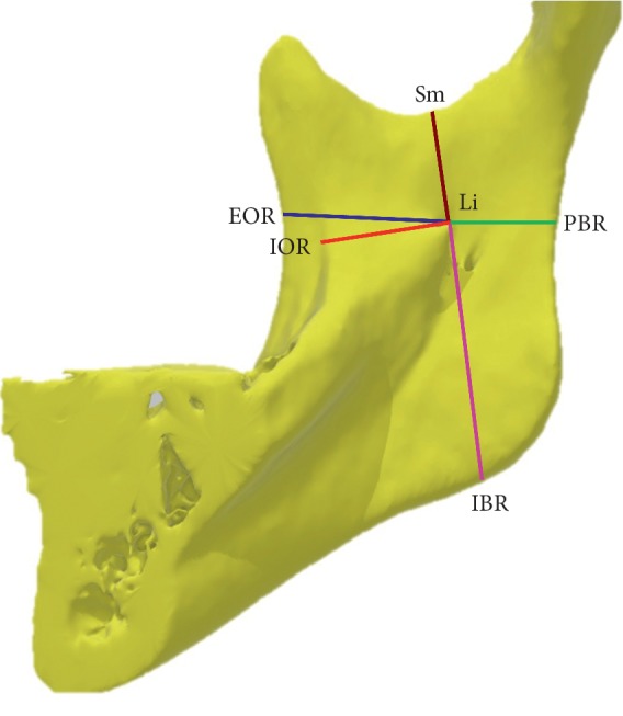
The study aimed to investigate and measure the anatomical relationship between the mandibular lingula (Li) and skeletal patterns using cone-beam computed tomography (CBCT). . In total, 72 participants (23 men and 49 women) were categorized into three groups according to their skeletal patterns (specifically, the A point-nasion-B point (ANB]) angle) as follows: Class I (0° < ANB < 4°), Class II (ANB ≥ 4°), and Class III (ANB ≤ 0°). The CBCT images of 144 rami were collected, and the distance from the Li to the external oblique ridge (Li-EOR), internal oblique ridge (Li-IOR), posterior border of the ramus (Li-PBR), inferior border of the ramus (Li-IBR), sigmoid notch (Li-Sm), and mandibular foramen (Li-MF) was examined. The Li-hMF (horizontal distance from the Li to the MF) and Li-vMF (vertical distance from the Li to the MF) were measured. The comparisons of gender, side (right and left), and skeletal patterns were then evaluated by statistical analysis.

RESULTS

The values of the Li-EOR and Li-PBR (19.99 mm and 15.93 mm, respectively) were significantly higher in men than in women (18.85 mm and 14.89 mm, respectively). Moreover, the Li-IBR was higher in men (32.91 mm) than in women (30.40 mm). Both sides (right and left) and skeletal patterns demonstrated that the Li-EOR, Li-IOR, and Li-PBR were not significantly different. Pearson's correlation test reported a strong correlation between the Li-EOR and Li-IOR (=0.610).

CONCLUSION

The distances from mandibula lingula to the external oblique ridge, posterior border of ramus, and inferior border of ramus were significantly longer in men than in women. Similarly, both horizontal and vertical distances from the lingula to the mandibular foramen were significantly longer in men than in women. Therefore, the results demonstrated that the Li was longer and more protruding in men than in women. With respect to the horizontal distance from the mandibular lingula to the mandibular foramen, of the three types of the skeletal system (Class I, Class II, and Class III), Class III was the significantly largest.

摘要

目的

本研究旨在通过锥形束 CT(CBCT)来探讨和测量下颌舌骨棘(Li)与骨骼形态之间的解剖关系。共有 72 名参与者(23 名男性和 49 名女性)根据其骨骼形态(具体为 A 点-鼻根点-B 点(ANB)角)分为三组:I 类(0°<ANB<4°)、II 类(ANB≥4°)和 III 类(ANB≤0°)。采集了 144 个支的 CBCT 图像,并检查了 Li 到外斜嵴(Li-EOR)、内斜嵴(Li-IOR)、下颌支后缘(Li-PBR)、下颌支下缘(Li-IBR)、乙状切迹(Li-Sm)和下颌孔(Li-MF)的距离。测量了 Li-hMF(Li 到 MF 的水平距离)和 Li-vMF(Li 到 MF 的垂直距离)。然后通过统计分析对性别、侧别(右侧和左侧)和骨骼形态进行了比较。

结果

男性的 Li-EOR 和 Li-PBR 值(19.99mm 和 15.93mm)明显高于女性(18.85mm 和 14.89mm)。此外,男性的 Li-IBR 更高(32.91mm),女性的 Li-IBR 更低(30.40mm)。左右两侧和骨骼形态均显示 Li-EOR、Li-IOR 和 Li-PBR 无显著差异。Pearson 相关检验显示 Li-EOR 和 Li-IOR 之间存在强相关(=0.610)。

结论

男性的下颌舌骨棘到外斜嵴、支后缘和支下缘的距离明显长于女性。同样,男性的 Li 到下颌孔的水平和垂直距离均明显长于女性。因此,结果表明男性的 Li 更长、更突出。就下颌舌骨棘到下颌孔的水平距离而言,在三种骨骼类型(I 类、II 类和 III 类)中,III 类明显最大。

https://cdn.ncbi.nlm.nih.gov/pmc/blobs/83ae/7042528/7cce82c13119/BMRI2020-2571534.002.jpg
https://cdn.ncbi.nlm.nih.gov/pmc/blobs/83ae/7042528/67b62494c98d/BMRI2020-2571534.001.jpg
https://cdn.ncbi.nlm.nih.gov/pmc/blobs/83ae/7042528/7cce82c13119/BMRI2020-2571534.002.jpg
https://cdn.ncbi.nlm.nih.gov/pmc/blobs/83ae/7042528/67b62494c98d/BMRI2020-2571534.001.jpg
https://cdn.ncbi.nlm.nih.gov/pmc/blobs/83ae/7042528/7cce82c13119/BMRI2020-2571534.002.jpg

相似文献

1
Dimension and Location of the Mandibular Lingula: Comparisons of Gender and Skeletal Patterns Using Cone-Beam Computed Tomography.下颌舌骨嵴的维度和位置:锥形束 CT 对性别和骨骼形态的比较。
Biomed Res Int. 2020 Feb 13;2020:2571534. doi: 10.1155/2020/2571534. eCollection 2020.
2
Three-dimensional evaluation of mandibular lingula: Comparisons of skeletal angle classifications and growth patterns.下颌舌隆突的三维评估:骨骼角度分类与生长模式的比较。
Orthod Craniofac Res. 2024 Jun;27(3):494-503. doi: 10.1111/ocr.12757. Epub 2024 Jan 21.
3
A detailed radiomorphometric analysis of the mandibular foramen, lingula and anti-lingula with a special emphasis on mandibular prognathism.下颌孔、下颌舌骨嵴和反舌骨嵴的详细放射形态分析,特别关注下颌前突。
J Stomatol Oral Maxillofac Surg. 2023 Feb;124(1):101260. doi: 10.1016/j.jormas.2022.08.003. Epub 2022 Aug 7.
4
Two-thirds anteroposterior ramus length is the preferred osteotomy point for intraoral vertical ramus osteotomy.三分之二的前向后弓长度是口内垂直下颌骨劈开术的首选截骨点。
Clin Oral Investig. 2022 Feb;26(2):1229-1239. doi: 10.1007/s00784-021-04094-1. Epub 2021 Jul 30.
5
The correlation between surgical reference points: antilingula, lingula, and mandibular foramen.手术参考点之间的相关性:反隆凸、隆凸和下颌孔。
J Stomatol Oral Maxillofac Surg. 2021 Dec;122(6):535-538. doi: 10.1016/j.jormas.2020.11.007. Epub 2020 Dec 8.
6
Relationship between the Mental Foramen, Mandibular Canal, and the Surgical Access Line of the Mandibular Posterior Teeth: A Cone-beam Computed Tomographic Analysis.下颌后牙的颏孔、下颌管与手术入路的关系:锥形束 CT 分析。
J Endod. 2017 Aug;43(8):1262-1266. doi: 10.1016/j.joen.2017.03.043. Epub 2017 Jun 22.
7
Measurement of mandibular lingula location using cone-beam computed tomography and internal oblique ridge-guided inferior alveolar nerve block.使用锥形束计算机断层扫描和内斜线引导下牙槽神经阻滞测量下颌小舌位置
J Korean Assoc Oral Maxillofac Surg. 2019 Jun;45(3):158-166. doi: 10.5125/jkaoms.2019.45.3.158. Epub 2019 Jun 28.
8
Determining the safety margin of mandibular lingula in sagittal split ramus osteotomy.确定下颌升支矢状劈开截骨术中下颌小舌的安全边界。
Surg Radiol Anat. 2024 Feb;46(2):159-166. doi: 10.1007/s00276-023-03291-9. Epub 2024 Jan 20.
9
Assessment of the Location of the Mandibular Lingula in Pediatric Patients Using Cone Beam Computed Tomography Images.使用锥形束计算机断层扫描图像评估儿科患者下颌小舌的位置
J Dent Child (Chic). 2018 May 15;85(2):58-65.
10
Correlation between the Mandibular Lingula Position and Some Anatomical Landmarks in Cone Beam CT.锥形束CT中下颌舌骨沟位置与一些解剖标志之间的相关性
Healthcare (Basel). 2021 Dec 17;9(12):1747. doi: 10.3390/healthcare9121747.

引用本文的文献

1
A Cone-Beam Computed Tomography Study of the Morphological and Morphometric Variations in the Mandibular Lingula and Its Clinical Implications.下颌小舌形态及形态计量学变异的锥形束计算机断层扫描研究及其临床意义
Diagnostics (Basel). 2025 Aug 18;15(16):2071. doi: 10.3390/diagnostics15162071.
2
Evaluation of positions of four lingula shapes for mandibular ramus surgery.下颌升支手术中四种舌形位置的评估。
Front Oral Health. 2025 Jan 7;5:1521227. doi: 10.3389/froh.2024.1521227. eCollection 2024.
3
Variations of the Lingula and Mandibular Ramus in the Context of Sagittal Split Ramus Osteotomy: A Cone Beam Computed Tomography Study Supporting an Ethnic-Centric Approach to Orthognathic Surgery.

本文引用的文献

1
Anatomical study of the location of the antilingula, lingula, and mandibular foramen for vertical ramus osteotomy.垂直下颌支截骨术中抗角部、角部及下颌孔位置的解剖学研究
Maxillofac Plast Reconstr Surg. 2018 Jul 25;40(1):15. doi: 10.1186/s40902-018-0155-3. eCollection 2018 Dec.
2
The variable position of the inferior alveolar nerve (IAN) in the mandibular ramus: a computed tomography (CT) study.下颌支中下牙槽神经(IAN)的可变位置:一项计算机断层扫描(CT)研究。
Surg Radiol Anat. 2018 Jun;40(6):653-665. doi: 10.1007/s00276-018-1973-9. Epub 2018 Jan 20.
3
Morphological evaluation of the mandibular lingula using cone-beam computed tomography.
矢状劈开下颌支截骨术中舌骨和下颌支的变异:一项锥形束计算机断层扫描研究,支持正颌外科以种族为中心的方法
Cureus. 2024 Aug 25;16(8):e67715. doi: 10.7759/cureus.67715. eCollection 2024 Aug.
4
Determining the safety margin of mandibular lingula in sagittal split ramus osteotomy.确定下颌升支矢状劈开截骨术中下颌小舌的安全边界。
Surg Radiol Anat. 2024 Feb;46(2):159-166. doi: 10.1007/s00276-023-03291-9. Epub 2024 Jan 20.
5
An Anatomical Study of Dry Mandibles to Determine the Important Surgical Reference Points in Ramus Osteotomy - An Evaluative Study.一项关于干燥下颌骨的解剖学研究,以确定下颌支截骨术中重要的手术参考点——一项评估性研究。
Ann Maxillofac Surg. 2023 Jan-Jun;13(1):9-12. doi: 10.4103/ams.ams_19_23. Epub 2023 Jun 30.
6
Morphological Study of the Mandibular Lingula and Antilingula by Cone-Beam Computed Tomography.锥形束计算机断层扫描对下颌小舌及小舌前区的形态学研究
Bioengineering (Basel). 2023 Jan 28;10(2):170. doi: 10.3390/bioengineering10020170.
7
Locating the Mandibular Lingula Using Cone-Beam Computed Tomography: A Literature Review.使用锥形束计算机断层扫描定位下颌舌骨嵴:文献综述
J Clin Med. 2023 Jan 22;12(3):881. doi: 10.3390/jcm12030881.
8
Association of Mandibular Posterior Anatomic Limit with Skeletal Patterns and Root Morphology Using Three-Dimensional Cone Beam Computed Tomography Comprehensive Analysis.使用三维锥形束计算机断层扫描综合分析下颌后部解剖学界限与骨骼形态及牙根形态的相关性
Diagnostics (Basel). 2022 Dec 2;12(12):3019. doi: 10.3390/diagnostics12123019.
9
Correlation between the Mandibular Lingula Position and Some Anatomical Landmarks in Cone Beam CT.锥形束CT中下颌舌骨沟位置与一些解剖标志之间的相关性
Healthcare (Basel). 2021 Dec 17;9(12):1747. doi: 10.3390/healthcare9121747.
10
Two-thirds anteroposterior ramus length is the preferred osteotomy point for intraoral vertical ramus osteotomy.三分之二的前向后弓长度是口内垂直下颌骨劈开术的首选截骨点。
Clin Oral Investig. 2022 Feb;26(2):1229-1239. doi: 10.1007/s00784-021-04094-1. Epub 2021 Jul 30.
使用锥形束计算机断层扫描对下颌小舌进行形态学评估。
Folia Morphol (Warsz). 2015;74(4):497-502. doi: 10.5603/FM.2015.0114.
4
Cone-beam computed tomography analysis of the shape, height, and location of the mandibular lingula.锥形束计算机断层扫描分析下颌小舌的形态、高度和位置。
Surg Radiol Anat. 2014 Mar;36(2):155-62. doi: 10.1007/s00276-013-1150-0. Epub 2013 Jun 23.
5
Anatomic study to determine a safe surgical reference point for mandibular ramus osteotomy.解剖学研究确定下颌支骨切开术的安全手术参考点。
J Craniomaxillofac Surg. 2014 Jan;42(1):22-7. doi: 10.1016/j.jcms.2013.01.041. Epub 2013 Feb 28.
6
Anatomic study of the mandibular foramen, lingula and antilingula in dry mandibles, and its statistical relationship between the true lingula and the antilingula.下颌孔、下颌舌骨嵴和反舌骨嵴的解剖学研究在干燥下颌骨中的应用,以及真舌骨和反舌骨之间的统计学关系。
Int J Oral Maxillofac Surg. 2012 Jan;41(1):74-8. doi: 10.1016/j.ijom.2011.08.009. Epub 2011 Sep 28.
7
The study of position of antilingula, midwaist of mandibular ramus and midpoint between coronoid process and gonion in relation to lingula of 92 Thai dried mandibles as potential surgical landmarks for vertical ramus osteotomy.对92具泰国干燥下颌骨的抗舌骨嵴位置、下颌支中腰部以及冠突与下颌角中点相对于舌骨嵴的位置进行研究,将其作为垂直下颌支截骨术潜在的手术标志。
Surg Radiol Anat. 2011 May;33(4):337-43. doi: 10.1007/s00276-010-0700-y. Epub 2010 Jul 16.
8
Shape, height, and location of the lingula for sagittal ramus osteotomy in Thais.泰国人矢状支截骨术中舌骨的形状、高度和位置。
Clin Anat. 2009 Oct;22(7):787-93. doi: 10.1002/ca.20849.
9
Accuracy of using the antilingula as a sole determinant of vertical ramus osteotomy position.将角前切迹作为垂直升支截骨术位置的唯一决定因素的准确性。
J Oral Maxillofac Surg. 2007 May;65(5):859-62. doi: 10.1016/j.joms.2006.05.065.
10
Esthetics and its relation to orthodontic therapy.美学及其与正畸治疗的关系。
Angle Orthod. 1950 Jul;20(3):168-78. doi: 10.1043/0003-3219(1950)020<0168:EAIRTO>2.0.CO;2.