• 文献检索
  • 文档翻译
  • 深度研究
  • 学术资讯
  • Suppr Zotero 插件Zotero 插件
  • 邀请有礼
  • 套餐&价格
  • 历史记录
应用&插件
Suppr Zotero 插件Zotero 插件浏览器插件Mac 客户端Windows 客户端微信小程序
定价
高级版会员购买积分包购买API积分包
服务
文献检索文档翻译深度研究API 文档MCP 服务
关于我们
关于 Suppr公司介绍联系我们用户协议隐私条款
关注我们

Suppr 超能文献

核心技术专利:CN118964589B侵权必究
粤ICP备2023148730 号-1Suppr @ 2026

文献检索

告别复杂PubMed语法,用中文像聊天一样搜索,搜遍4000万医学文献。AI智能推荐,让科研检索更轻松。

立即免费搜索

文件翻译

保留排版,准确专业,支持PDF/Word/PPT等文件格式,支持 12+语言互译。

免费翻译文档

深度研究

AI帮你快速写综述,25分钟生成高质量综述,智能提取关键信息,辅助科研写作。

立即免费体验

一种用于研究和临床实践中牙根及根管系统分类的新系统的优势与应用

Advantages and Applications of a New System for Classifying Roots and Canal Systems in Research and Clinical Practice.

作者信息

Ahmed Hany Mohamed Aly, Dummer Paul Michael Howell

机构信息

Department of Restorative Dentistry, University of Malaya School of Dentistry, Kuala Lumpur, Malaysia.

School of Dentistry, College of Biomedical and Life Sciences, Cardiff University, Cardiff, UK.

出版信息

Eur Endod J. 2017 Dec 21;3(1):9-17. doi: 10.5152/eej.2017.17064. eCollection 2018.

DOI:10.5152/eej.2017.17064
PMID:32161850
原文链接:https://pmc.ncbi.nlm.nih.gov/articles/PMC7024718/
Abstract

OBJECTIVE

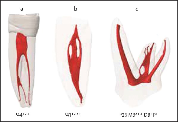
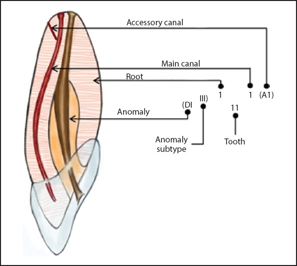
A new coding system for classifying the roots, main and accessory canals as well as developmental anomalies has been introduced recently. This paper discusses the advantages and potential application of the new system in research and clinical practice.

METHODS

A comprehensive analysis was undertaken on the most common, existing classification for root canal morphology. The advantages and potential applications of a new system for classifying roots and canal systems in research and clinical practice are discussed.

RESULTS

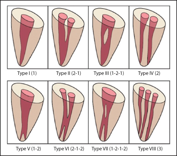
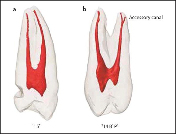
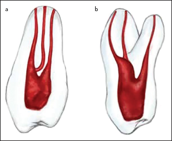
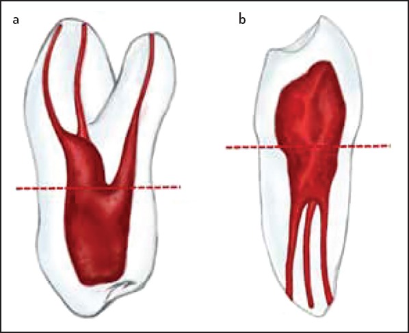
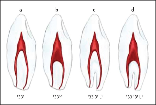
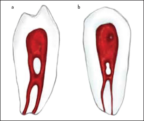
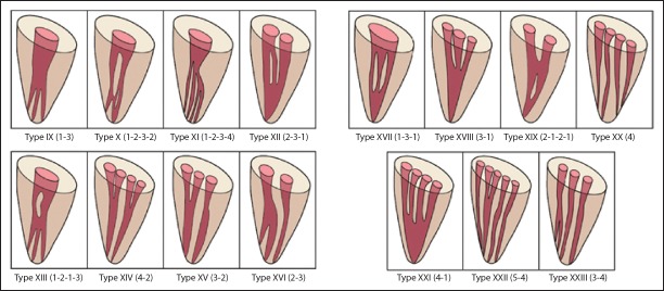
The analysis demonstrates deficiencies of the existing classification including lack of information on the number of roots, pulp chamber outline, lack of clarity in multi-rooted teeth, inability to define complex root canal configurations. The new coding system addresses the root and canal morphology in an accurate and systematic manner to provide detailed information of the tooth, root and canal anatomical features.

CONCLUSION

With current advances in endodontic research and practice and the increasing body of knowledge on root and canal morphology, the deficiencies of the existing system used for classifying root canal morphology have become more apparent. The new system for classifying root, main and accessory canal morphology as well as teeth with anomalies has the potential to be used in research, clinical practice and education to accurately reflect the real anatomy of a tooth.

摘要

目的

最近引入了一种用于对牙根、主根管和副根管以及发育异常进行分类的新编码系统。本文讨论了该新系统在研究和临床实践中的优势及潜在应用。

方法

对最常见的现有根管形态分类进行了全面分析。讨论了一种用于在研究和临床实践中对牙根和根管系统进行分类的新系统的优势及潜在应用。

结果

分析表明现有分类存在缺陷,包括缺乏关于牙根数量、髓腔轮廓的信息,多根牙情况不清晰,无法定义复杂的根管形态。新编码系统以准确且系统的方式处理牙根和根管形态,以提供牙齿、牙根和根管解剖特征的详细信息。

结论

随着当前牙髓病研究和实践的进展以及关于牙根和根管形态的知识不断增加,用于分类根管形态的现有系统的缺陷变得更加明显。用于对牙根、主根管和副根管形态以及异常牙齿进行分类的新系统有潜力用于研究、临床实践和教育,以准确反映牙齿的真实解剖结构。

https://cdn.ncbi.nlm.nih.gov/pmc/blobs/449d/7024718/5f92a1f2efb7/EEJ-3-9-g011.jpg
https://cdn.ncbi.nlm.nih.gov/pmc/blobs/449d/7024718/3b2fee87e57f/EEJ-3-9-g001.jpg
https://cdn.ncbi.nlm.nih.gov/pmc/blobs/449d/7024718/34e510c6d15a/EEJ-3-9-g002.jpg
https://cdn.ncbi.nlm.nih.gov/pmc/blobs/449d/7024718/ec540fc94421/EEJ-3-9-g003.jpg
https://cdn.ncbi.nlm.nih.gov/pmc/blobs/449d/7024718/180ee3af98b9/EEJ-3-9-g004.jpg
https://cdn.ncbi.nlm.nih.gov/pmc/blobs/449d/7024718/bb007a1f4501/EEJ-3-9-g005.jpg
https://cdn.ncbi.nlm.nih.gov/pmc/blobs/449d/7024718/a1a92fe6632a/EEJ-3-9-g006.jpg
https://cdn.ncbi.nlm.nih.gov/pmc/blobs/449d/7024718/53158cebeb95/EEJ-3-9-g007.jpg
https://cdn.ncbi.nlm.nih.gov/pmc/blobs/449d/7024718/592569c46346/EEJ-3-9-g008.jpg
https://cdn.ncbi.nlm.nih.gov/pmc/blobs/449d/7024718/918ecfed70db/EEJ-3-9-g009.jpg
https://cdn.ncbi.nlm.nih.gov/pmc/blobs/449d/7024718/586b5ac6b5ee/EEJ-3-9-g010.jpg
https://cdn.ncbi.nlm.nih.gov/pmc/blobs/449d/7024718/5f92a1f2efb7/EEJ-3-9-g011.jpg

相似文献

1
Advantages and Applications of a New System for Classifying Roots and Canal Systems in Research and Clinical Practice.一种用于研究和临床实践中牙根及根管系统分类的新系统的优势与应用
Eur Endod J. 2017 Dec 21;3(1):9-17. doi: 10.5152/eej.2017.17064. eCollection 2018.
2
A new system for classifying tooth, root and canal anomalies.一种新的牙齿、牙根和根管畸形分类系统。
Int Endod J. 2018 Apr;51(4):389-404. doi: 10.1111/iej.12867. Epub 2017 Nov 1.
3
Application of a new system for classifying root and canal anatomy in studies involving micro-computed tomography and cone beam computed tomography: Explanation and elaboration.一种新的根管解剖分类系统在涉及微计算机断层扫描和锥形束计算机断层扫描研究中的应用:说明和阐述。
Int Endod J. 2021 Jul;54(7):1056-1082. doi: 10.1111/iej.13486. Epub 2021 Apr 18.
4
Application of a new system for classifying tooth, root and canal morphology in the primary dentition.应用一种新的系统对乳恒牙列的牙、根和根管形态进行分类。
Int Endod J. 2020 Jan;53(1):27-35. doi: 10.1111/iej.13199. Epub 2019 Oct 1.
5
Application of a new System for Classifying Root and Canal Anatomy in Clinical Practice - Explanation and Elaboration.应用根管解剖分类新系统于临床实践:说明与详述。
Eur Endod J. 2021 Aug;6(2):132-142. doi: 10.14744/eej.2021.38257.
6
A new system for classifying root and root canal morphology.一种新的根管形态分类系统。
Int Endod J. 2017 Aug;50(8):761-770. doi: 10.1111/iej.12685. Epub 2016 Oct 17.
7
A new system for classifying accessory canal morphology.一种新的副根管形态分类系统。
Int Endod J. 2018 Feb;51(2):164-176. doi: 10.1111/iej.12800. Epub 2017 Jul 31.
8
Implications of root, pulp chamber, and canal anatomy on pulpotomy and revitalization procedures.根管、牙髓腔和根管解剖学对活髓切断术和牙髓再生术的影响。
Clin Oral Investig. 2023 Nov;27(11):6357-6369. doi: 10.1007/s00784-023-05284-9. Epub 2023 Oct 23.
9
Root and Root Canal Morphology Classification Systems.牙根及根管形态分类系统
Int J Dent. 2021 Feb 19;2021:6682189. doi: 10.1155/2021/6682189. eCollection 2021.
10
Root and canal morphology of maxillary premolar teeth in an Egyptian subpopulation using two classification systems: a cone beam computed tomography study.采用两种分类系统对埃及亚人群上颌前磨牙的根管和牙根形态进行的锥形束 CT 研究。
Int Endod J. 2019 Mar;52(3):267-278. doi: 10.1111/iej.13016. Epub 2018 Nov 8.

引用本文的文献

1
Assessment of root and root canal morphology in maxillary molars with fused roots using Cone Beam Computer Tomography (CBCT) in a Sri Lankan population.在斯里兰卡人群中使用锥形束计算机断层扫描(CBCT)评估融合根上颌磨牙的牙根及根管形态。
J Oral Biol Craniofac Res. 2025 Nov-Dec;15(6):1297-1304. doi: 10.1016/j.jobcr.2025.08.016. Epub 2025 Aug 19.
2
Micro-computed tomographic evaluation of root and canal anatomy of maxillary first premolars in Iraqi sub-population.伊拉克亚人群上颌第一前磨牙牙根及根管解剖结构的显微计算机断层扫描评估
Sci Rep. 2025 Mar 28;15(1):10821. doi: 10.1038/s41598-025-94590-1.
3
Prevalence of Three-Rooted Mandibular First Molars Among Jordanian Royal Medical Services Patients Using Cone-Beam Computed Tomography.

本文引用的文献

1
Endodontic Management of a Mandibular First Molar with Unusual Canal Morphology.下颌第一磨牙根管形态异常的牙髓治疗
Eur Endod J. 2017 Feb 16;2(1):1-4. doi: 10.5152/eej.2017.17042. eCollection 2017.
2
A new system for classifying tooth, root and canal anomalies.一种新的牙齿、牙根和根管畸形分类系统。
Int Endod J. 2018 Apr;51(4):389-404. doi: 10.1111/iej.12867. Epub 2017 Nov 1.
3
A new system for classifying accessory canal morphology.一种新的副根管形态分类系统。
约旦皇家医疗服务机构患者中使用锥形束计算机断层扫描技术检测到的下颌第一磨牙三根发生率
Cureus. 2024 Nov 15;16(11):e73747. doi: 10.7759/cureus.73747. eCollection 2024 Nov.
4
Evaluation of root canal morphology of mandibular premolars in Pakistani population using the new classification: a CBCT study.利用新分类评估巴基斯坦人群下颌前磨牙根管形态:CBCT 研究。
BMC Oral Health. 2024 Nov 20;24(1):1414. doi: 10.1186/s12903-024-05149-x.
5
A New Methodology to Determine the Orifice for Root Canal Configurations in First Permanent Molar Root and Canal Morphologies Using Micro-Computed Tomography.一种使用微型计算机断层扫描确定第一恒磨牙根管形态根管口的新方法
J Clin Med. 2023 Dec 22;13(1):71. doi: 10.3390/jcm13010071.
6
Characterization of the root and canal anatomy of maxillary premolar teeth in an Iraqi subpopulation: a cone beam computed tomography study.伊拉克亚人群上颌前磨牙的根管解剖结构特征:锥形束 CT 研究。
Odontology. 2024 Apr;112(2):570-587. doi: 10.1007/s10266-023-00870-5. Epub 2023 Nov 13.
7
Evaluation of the Frequency and Anatomy of Radix Entomolaris and Paramolaris in Lower Molars by Cone Beam Computed Tomography (CBCT) in Northern Iran, 2020-2021: A Retrospective Study.2020 - 2021年伊朗北部锥形束计算机断层扫描(CBCT)评估下颌磨牙远中根及额外根的频率和解剖结构:一项回顾性研究
Cureus. 2023 Oct 11;15(10):e46854. doi: 10.7759/cureus.46854. eCollection 2023 Oct.
8
Revisiting Premolars Using Cone-Beam Computed Tomography Analysis and Classifying Their Roots and Root Canal Morphology Using Newer Classification.使用锥形束计算机断层扫描分析重新审视前磨牙,并使用更新的分类方法对其牙根和根管形态进行分类。
Cureus. 2023 May 6;15(5):e38623. doi: 10.7759/cureus.38623. eCollection 2023 May.
9
A microcomputed tomographic analysis of the morphological variabilities and incidence of extra canals in mandibular first molar teeth in an Egyptian subpopulation.埃及人群下颌第一磨牙形态变异和额外管发生率的显微 CT 分析。
Sci Rep. 2023 Jun 2;13(1):8985. doi: 10.1038/s41598-023-36005-7.
10
Evaluation of root and canal morphology of mandibular premolar amongst Saudi subpopulation using the new system of classification: a CBCT study.应用新分类系统评估沙特人群下颌前磨牙的根管及牙根形态:CBCT 研究。
BMC Oral Health. 2023 May 15;23(1):291. doi: 10.1186/s12903-023-03002-1.
Int Endod J. 2018 Feb;51(2):164-176. doi: 10.1111/iej.12800. Epub 2017 Jul 31.
4
Root and root canal morphology of the permanent dentition in a Caucasian population: a cone-beam computed tomography study.高加索人群恒牙的根管和根管形态:锥形束 CT 研究。
Int Endod J. 2017 Nov;50(11):1013-1026. doi: 10.1111/iej.12724. Epub 2017 Jan 3.
5
A new system for classifying root and root canal morphology.一种新的根管形态分类系统。
Int Endod J. 2017 Aug;50(8):761-770. doi: 10.1111/iej.12685. Epub 2016 Oct 17.
6
Micro-Computed Tomography Assessment of Apical Accessory Canal Morphologies.根尖副根管形态的微计算机断层扫描评估
J Endod. 2016 May;42(5):798-802. doi: 10.1016/j.joen.2016.02.006. Epub 2016 Mar 11.
7
Micro-computed tomographic analysis of the root canal morphology of the distal root of mandibular first molar.下颌第一磨牙远中根根管形态的显微计算机断层扫描分析
J Endod. 2015 Feb;41(2):231-6. doi: 10.1016/j.joen.2014.09.024. Epub 2014 Nov 12.
8
Three root canals in the mesiobuccal root of maxillary molars: case reports and literature review.上颌磨牙近中颊根中的三根根管:病例报告与文献综述
J Endod. 2014 Dec;40(12):2087-94. doi: 10.1016/j.joen.2014.07.034. Epub 2014 Oct 16.
9
Prevalence of middle mesial canals in mandibular molars after guided troughing under high magnification: an in vivo investigation.高倍放大引导下磨开后下颌磨牙近中中根管的患病率:一项体内研究
J Endod. 2015 Feb;41(2):164-8. doi: 10.1016/j.joen.2014.09.013. Epub 2014 Oct 30.
10
Root canal treatment of a maxillary second premolar with two palatal roots: A case report.上颌第二前磨牙双腭根的根管治疗:一例报告
J Conserv Dent. 2014 May;17(3):290-2. doi: 10.4103/0972-0707.131807.