Sugasawa Takehito, Kuji Tomoaki, Aoki Kai, Yanazawa Koki, Takenouchi Akiko, Watanabe Makoto, Tome Yoshiya, Takeuchi Yoshinori, Aita Yuichi, Yahagi Naoya, Shishikura Yasuhiro, Ono Seiko, Yoshida Yasuko, Kawakami Yasushi, Takekoshi Kazuhiro
Laboratory of Laboratory/Sports Medicine, Division of Clinical Medicine, Faculty of Medicine, University of Tsukuba, 1-1-1 Tennodai, Tsukuba, Ibaraki 305-8577, Japan.
Doctoral Program in Sports Medicine, Graduate School of Comprehensive Human Sciences, University of Tsukuba, 1-1-1 Tennodai, Tsukuba, Ibaraki 305-8577, Japan.
Biomedicines. 2020 Mar 10;8(3):56. doi: 10.3390/biomedicines8030056.
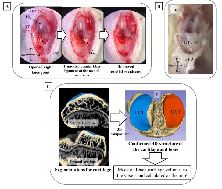
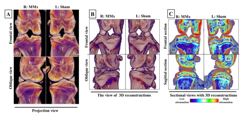
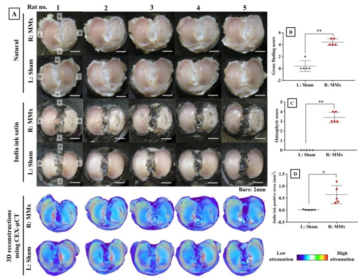
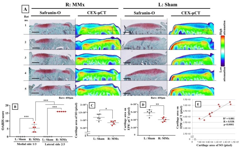
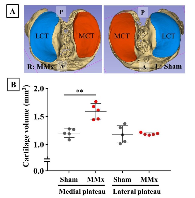
The aim of this study was to clarify degradation characteristics in each tissue of the knee complex of a medial meniscectomy (MMx)-induced knee osteoarthritis (KOA) animal model using classical methods and an alternative comprehensive evaluation method called contrast-enhanced X-ray micro-computed tomography (CEX-μCT), which was developed in the study. Surgical MMx was performed in the right knee joints of five male Wistar rats to induce KOA. At four weeks post-surgery, the synovitis was evaluated using quantitative polymerase chain reaction (qPCR). Degradations of the articular cartilage of the tibial plateau were evaluated using classical methods and CEX-μCT. Evaluation of the synovitis demonstrated significantly increased expression levels of inflammation-associated marker genes in MMx-treated knees compared with those in sham-treated knees. Evaluation of the articular cartilage using classical methods showed that MMx fully induced degradation of the cartilage. Evaluation using CEX-μCT showed that local areas of the medial cartilage of the tibial plateau were significantly reduced in MMx-treated knees compared with those in sham-treated knees. On the other hand, total cartilage volumes were significantly increased in MMx-treated knees. On the basis of the findings of this study, the method could be relevant to study new treatments in KOA research.
本研究的目的是使用经典方法以及一种在本研究中开发的名为对比增强X射线显微计算机断层扫描(CEX-μCT)的替代综合评估方法,阐明内侧半月板切除术(MMx)诱导的膝骨关节炎(KOA)动物模型膝关节复合体各组织中的退变特征。对五只雄性Wistar大鼠的右膝关节进行手术MMx以诱导KOA。术后四周,使用定量聚合酶链反应(qPCR)评估滑膜炎。使用经典方法和CEX-μCT评估胫骨平台关节软骨的退变情况。滑膜炎评估显示,与假手术组相比,MMx处理组膝关节中炎症相关标记基因的表达水平显著升高。使用经典方法对关节软骨进行评估表明,MMx完全诱导了软骨退变。使用CEX-μCT评估显示,与假手术组相比,MMx处理组膝关节胫骨平台内侧软骨的局部区域显著减少。另一方面,MMx处理组膝关节的软骨总体积显著增加。基于本研究的结果,该方法可能与KOA研究中的新治疗方法相关。