Cai Heguo, Qu Ning, Chen Xiaolei, Zhou Yang, Zheng Xinpeng, Zhang Bing, Xia Chun
Zhongshan Hospital, Xiamen University, Fujian 361004, China.
The Third Hospital of Xiamen, Fujian, China, Fujian 361000, China.
Oncotarget. 2017 Dec 15;9(4):4461-4474. doi: 10.18632/oncotarget.23286. eCollection 2018 Jan 12.
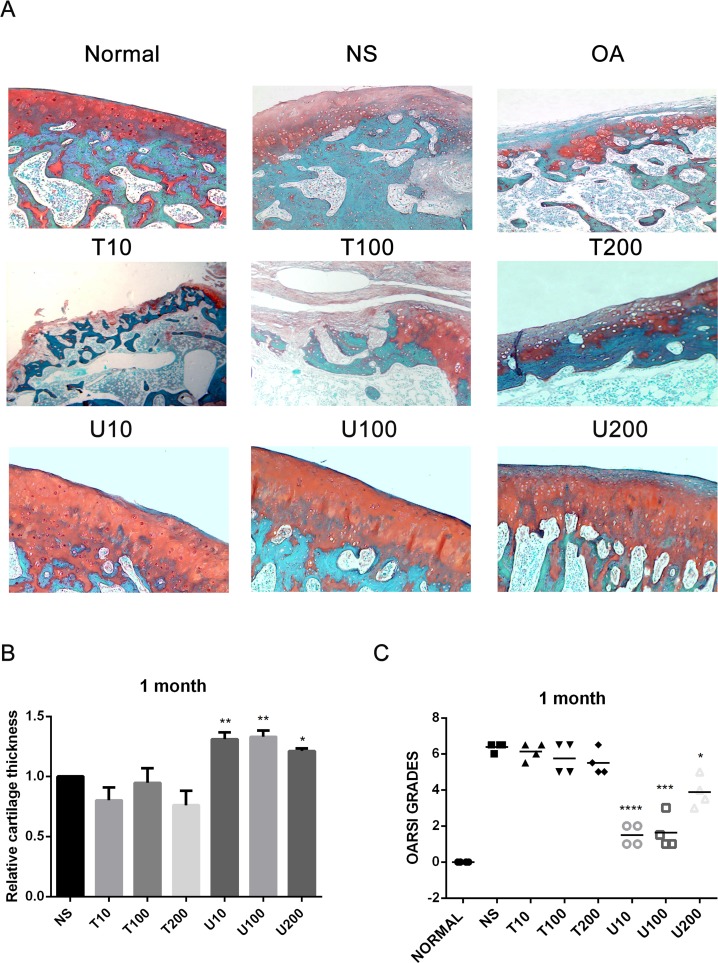
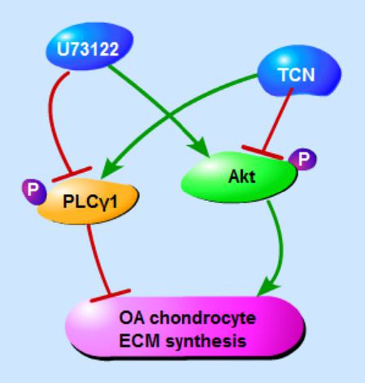
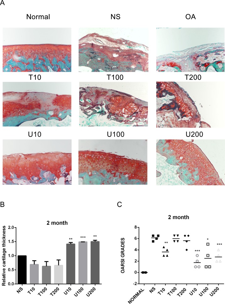
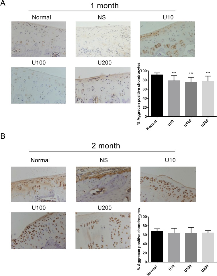
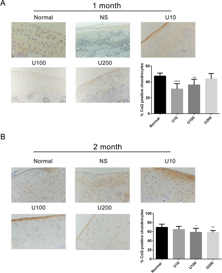
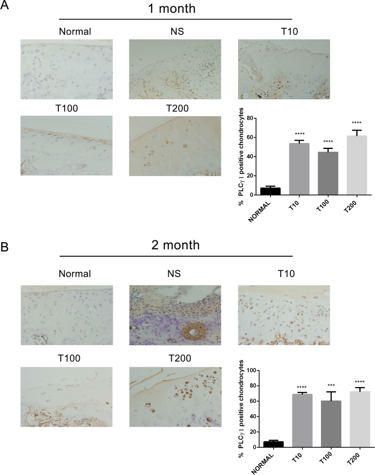
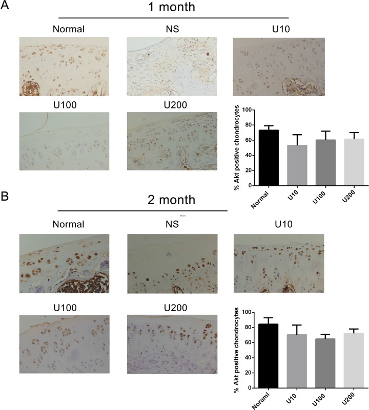
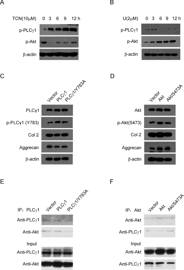
Previous studies have addressed the involvement of phosphoinositide-specifc phospholipase γ1 (PLCγ1) and protein kinase B (PKB/Akt) in osteoarthritis (OA) pathogenesis, but it is not ascertained the possibility of them to be potential targets for OA therapy. Here, through local intra-articular injection of PLCγ or Akt inhibitor in a rat OA model induced by anterior cruciate ligament transaction plus medial meniscus resection, the architecture of chondrocyte and matrix organization of articular cartilage were observed using histopathological assays and Aggrecan, Col2, PLCγ1, and Akt levels were detected using immunohistochemistry assays. By treatment of Akt or PLCγ inhibitor and transfection of different PLCγ1- or Akt-expressing vectors in rat OA model chondrocytes, Aggrecan, Col2, PLCγ1, p-PLCγ1, Akt, and p-Akt levels were detected using western blotting analysis. The binding between PLCγ1 and Akt was assessed with co-immunoprecipitation assays in human OA chondrocytes. These results showed that PLCγ inhibition protected chondrocytes against OA, but Akt inhibition did not dramatically aggravate OA progression. There were mutual antagonism and binding between PLCγ1 and Akt that could be regulated by their phosphorylation levels. Consequently, the data reveal that the inhibition of PLCγ1 may provide an attractive therapeutic target for OA therapy, implicating its binding to Akt.
先前的研究已经探讨了磷酸肌醇特异性磷脂酶γ1(PLCγ1)和蛋白激酶B(PKB/Akt)在骨关节炎(OA)发病机制中的作用,但尚未确定它们作为OA治疗潜在靶点的可能性。在此,通过在由前交叉韧带切断加内侧半月板切除诱导的大鼠OA模型中局部关节内注射PLCγ或Akt抑制剂,使用组织病理学分析观察软骨细胞的结构和关节软骨的基质组织,并使用免疫组织化学分析检测聚集蛋白聚糖、Ⅱ型胶原、PLCγ1和Akt水平。通过在大鼠OA模型软骨细胞中用Akt或PLCγ抑制剂处理并转染不同的PLCγ1或Akt表达载体,使用蛋白质印迹分析检测聚集蛋白聚糖、Ⅱ型胶原、PLCγ1、磷酸化PLCγ1、Akt和磷酸化Akt水平。在人OA软骨细胞中用免疫共沉淀分析评估PLCγ1与Akt之间的结合。这些结果表明,抑制PLCγ可保护软骨细胞免受OA影响,但抑制Akt不会显著加重OA进展。PLCγ1与Akt之间存在相互拮抗和结合,且这种关系可由它们的磷酸化水平调节。因此,数据表明抑制PLCγ1可能为OA治疗提供一个有吸引力的治疗靶点,这与其与Akt的结合有关。