Goyeneche Alicia, Lisio Michael-Anthony, Fu Lili, Srinivasan Rekha, Valdez Capuccino Juan, Gao Zu-Hua, Telleria Carlos
Experimental Pathology Unit, Department of Pathology, Faculty of Medicine, McGill University, Montreal, QC H3A 2B4, Canada.
Division of Basic Biomedical Sciences, Sanford School of Medicine, The University of South Dakota, Vermillion, SD 57069, USA.
Cancers (Basel). 2020 Mar 16;12(3):699. doi: 10.3390/cancers12030699.
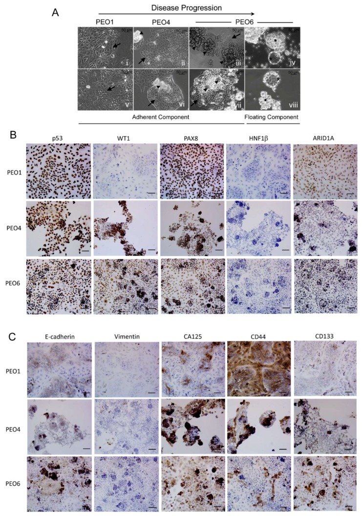
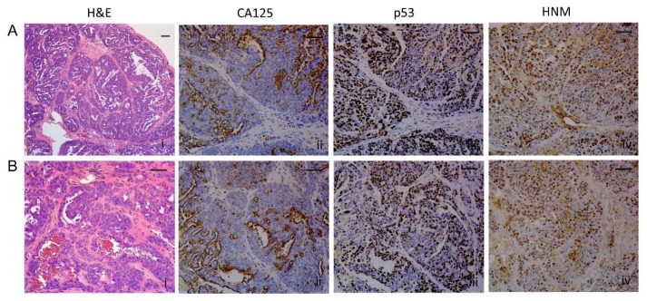
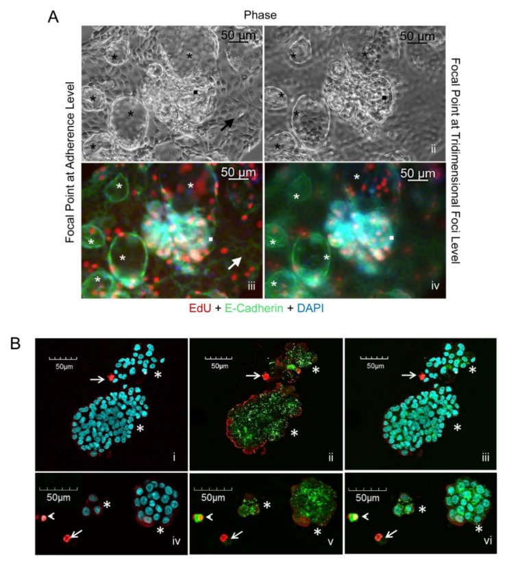
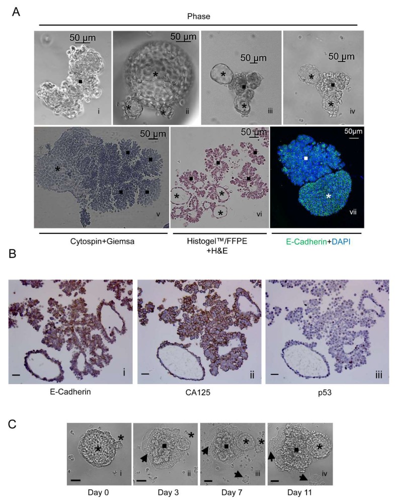
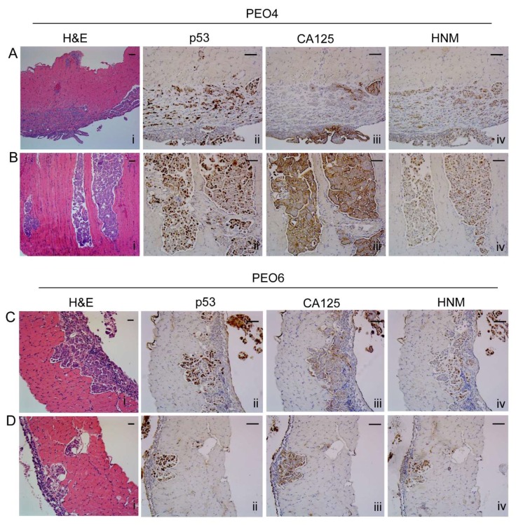
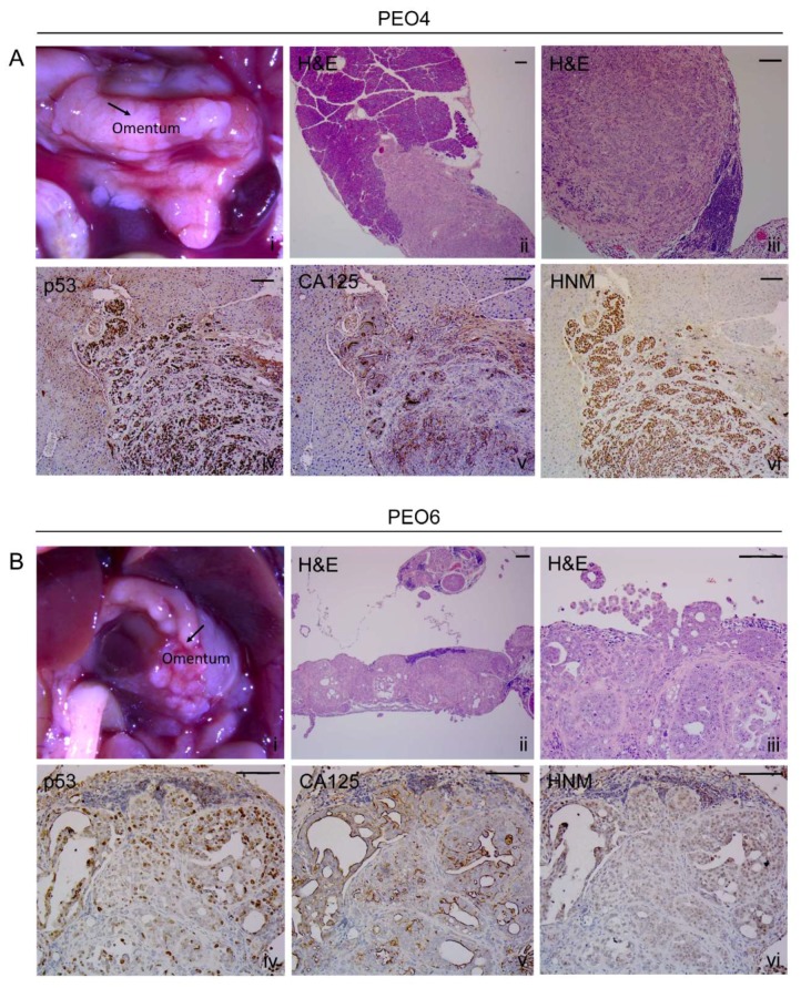
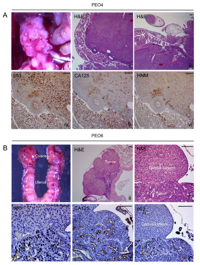
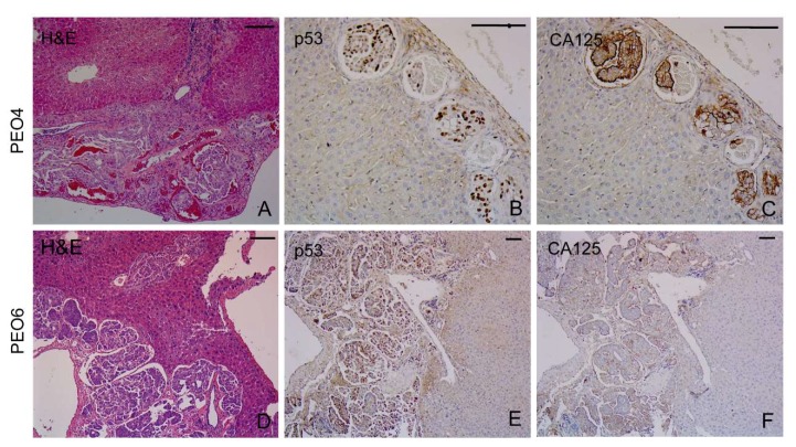
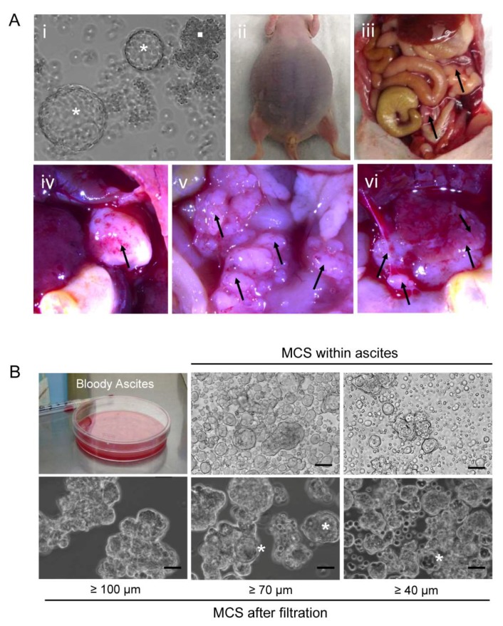
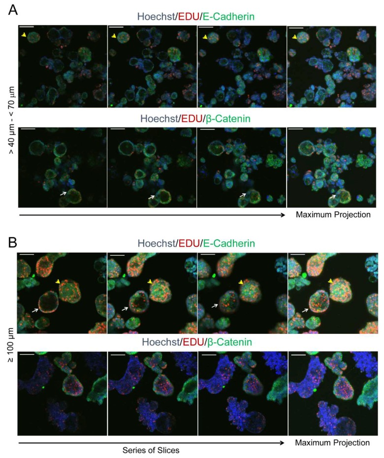
Many studies have examined the biology, genetics, and chemotherapeutic response of ovarian cancer's solid component; its liquid facet, however, remains critically underinvestigated. Floating within peritoneal effusions known as ascites, ovarian cancer cells form multicellular structures, creating a cancer niche in suspension. This study explores the pathobiology of spontaneously formed, multicellular, ovarian cancer structures derived from serous ovarian cancer cells isolated along disease evolution. It also tests their capacity to cause peritoneal disease in immunosuppressed mice. Results stem from an analysis of cell lines representing the most frequently diagnosed ovarian cancer histotype (high-grade serous ovarian cancer), derived from ascites of the same patient at distinct stages of disease progression. When cultured under adherent conditions, in addition to forming cellular monolayers, the cultures developed areas in which the cells grew upwards, forming densely packed multilayers that ultimately detached from the bottom of the plates and lived as free-floating, multicellular structures. The capacity to form foci and to develop multicellular structures was proportional to disease progression at the time of ascites extraction. Self-assembled in culture, these structures varied in size, were either compact or hollow, irregular, or spheroidal, and exhibited replicative capacity and an epithelial nature. Furthermore, they fully recreated ovarian cancer disease in immunosuppressed mice: accumulation of malignant ascites and pleural effusions; formation of discrete, solid, macroscopic, peritoneal tumors; and microscopic growths in abdominal organs. They also reproduced the histopathological features characteristic of high-grade serous ovarian cancer when diagnosed in patients. The following results encourage the development of therapeutic interventions to interrupt the formation and/or survival of multicellular structures that constitute a floating niche in the peritoneal fluid, which in turn halts disease progression and prevents recurrence.
许多研究都对卵巢癌实体成分的生物学、遗传学及化疗反应进行了探究;然而,其液体部分仍严重缺乏研究。卵巢癌细胞漂浮在被称为腹水的腹腔积液中,形成多细胞结构,在悬浮状态下营造出一个癌症微环境。本研究探索了源自沿疾病进展过程分离出的浆液性卵巢癌细胞的自发形成的多细胞卵巢癌结构的病理生物学。同时还测试了这些结构在免疫抑制小鼠中引发腹膜疾病的能力。研究结果源于对代表最常见诊断的卵巢癌组织类型(高级别浆液性卵巢癌)的细胞系的分析,这些细胞系取自同一患者在疾病进展不同阶段的腹水。在贴壁条件下培养时,这些细胞系除了形成细胞单层外,还形成了细胞向上生长的区域,形成紧密堆积的多层结构,最终从培养皿底部脱离,以自由漂浮的多细胞结构形式存在。形成病灶和发展多细胞结构的能力与抽取腹水时的疾病进展程度成正比。这些结构在培养中自行组装,大小各异,有致密的或中空的、不规则的或球形的,具有复制能力且呈上皮性质。此外,它们在免疫抑制小鼠中完全重现了卵巢癌疾病:恶性腹水和胸腔积液的积聚;离散的、实性的、肉眼可见的腹膜肿瘤的形成;以及腹部器官中的微观生长。当在患者中诊断时,它们还重现了高级别浆液性卵巢癌的组织病理学特征。以下研究结果促使人们开发治疗干预措施,以阻断构成腹膜液中漂浮微环境的多细胞结构的形成和/或存活,进而阻止疾病进展并预防复发。