Dana-Farber Cancer Institute, 450 Brookline Avenue, Boston, MA, 02215, USA.
Harvard Medical School, 25 Shattuck Street, Boston, MA, 02115, USA.
Nat Commun. 2020 Mar 19;11(1):1459. doi: 10.1038/s41467-020-15315-8.
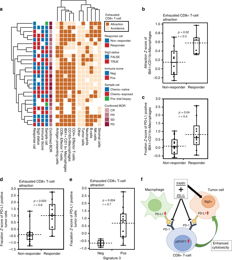
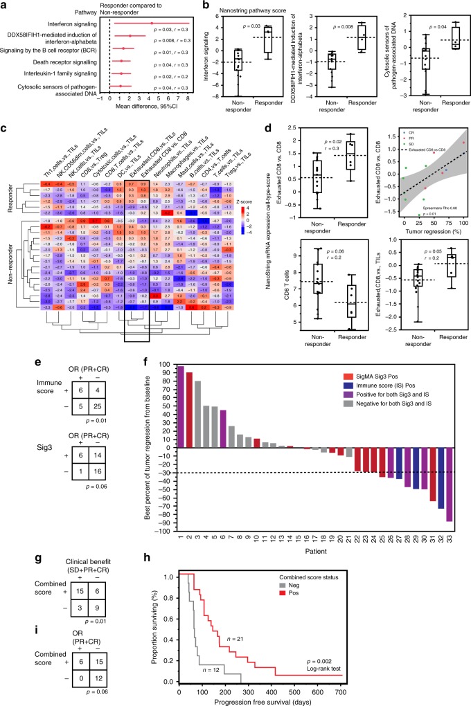
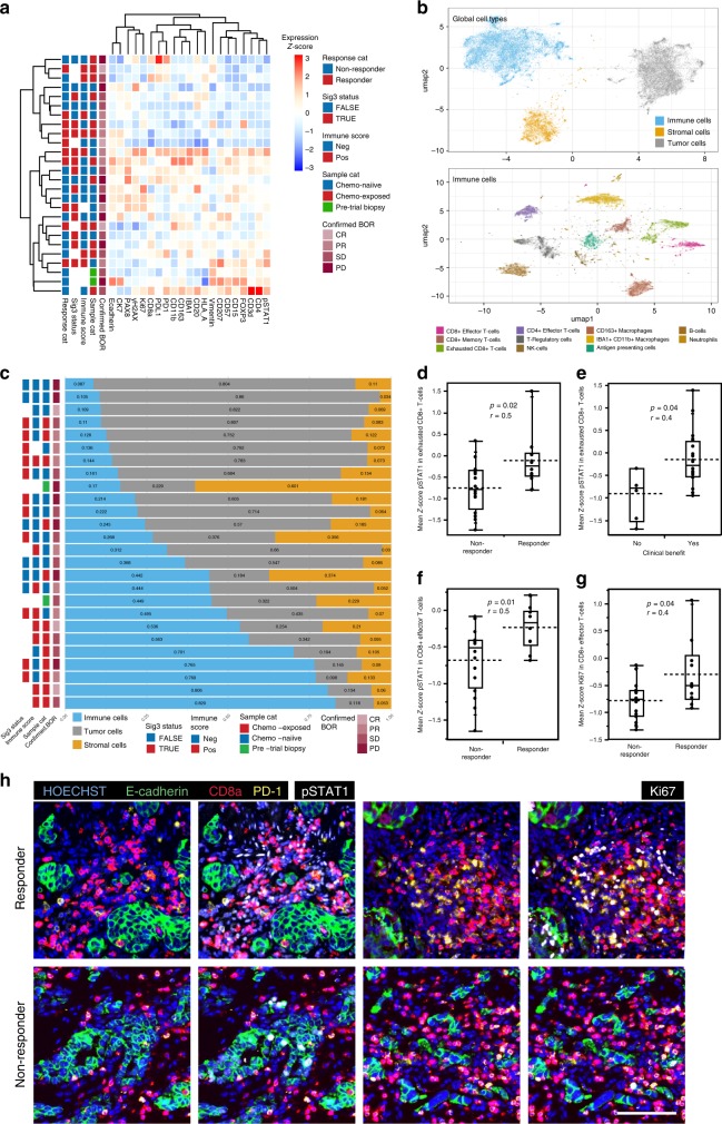
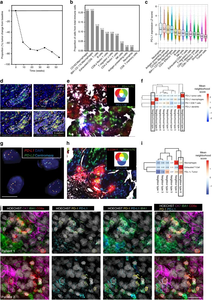
Combined PARP and immune checkpoint inhibition has yielded encouraging results in ovarian cancer, but predictive biomarkers are lacking. We performed immunogenomic profiling and highly multiplexed single-cell imaging on tumor samples from patients enrolled in a Phase I/II trial of niraparib and pembrolizumab in ovarian cancer (NCT02657889). We identify two determinants of response; mutational signature 3 reflecting defective homologous recombination DNA repair, and positive immune score as a surrogate of interferon-primed exhausted CD8 + T-cells in the tumor microenvironment. Presence of one or both features associates with an improved outcome while concurrent absence yields no responses. Single-cell spatial analysis reveals prominent interactions of exhausted CD8 + T-cells and PD-L1 + macrophages and PD-L1 + tumor cells as mechanistic determinants of response. Furthermore, spatial analysis of two extreme responders shows differential clustering of exhausted CD8 + T-cells with PD-L1 + macrophages in the first, and exhausted CD8 + T-cells with cancer cells harboring genomic PD-L1 and PD-L2 amplification in the second.
聚 PARP 和免疫检查点抑制在卵巢癌中已取得令人鼓舞的结果,但缺乏预测性生物标志物。我们对在卵巢癌的 niraparib 和 pembrolizumab 的 I/II 期试验(NCT02657889)中入组的患者的肿瘤样本进行了免疫基因组分析和高度多重化的单细胞成像。我们确定了两种反应决定因素;反映同源重组 DNA 修复缺陷的突变特征 3,以及肿瘤微环境中干扰素引发的耗尽 CD8+T 细胞的阳性免疫评分作为替代物。存在一个或两个特征与改善的结果相关,而同时缺乏则没有反应。单细胞空间分析揭示了耗尽的 CD8+T 细胞和 PD-L1+巨噬细胞以及 PD-L1+肿瘤细胞之间的突出相互作用,作为反应的机制决定因素。此外,对两个极端反应者的空间分析表明,在第一个反应者中,耗尽的 CD8+T 细胞与 PD-L1+巨噬细胞的聚类不同,而在第二个反应者中,耗尽的 CD8+T 细胞与携带基因组 PD-L1 和 PD-L2 扩增的癌细胞聚类不同。