Institute of Translational Medicine, Faculty of Health Sciences, University of Macau, Taipa, Macao SAR, China.
Institute of Chinese Medical Sciences, University of Macau, Taipa, Macao SAR, China.
Theranostics. 2020 Feb 3;10(7):2897-2917. doi: 10.7150/thno.40495. eCollection 2020.
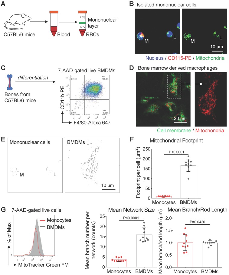
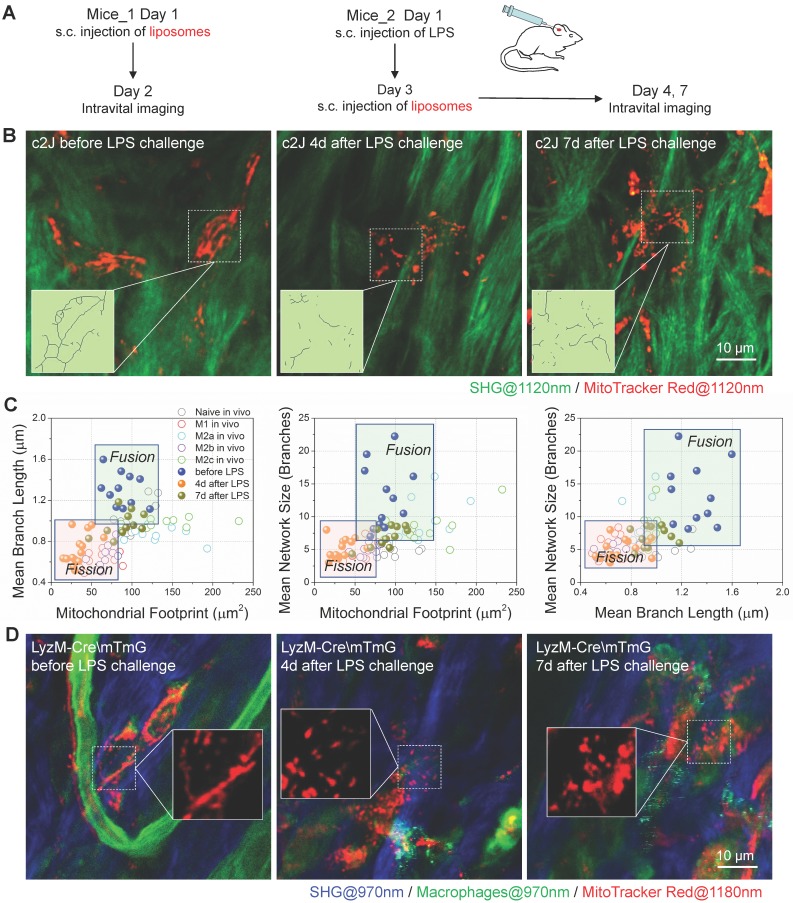
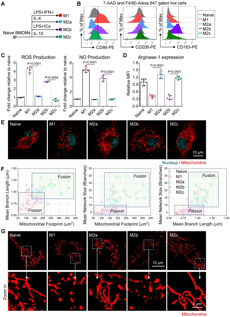
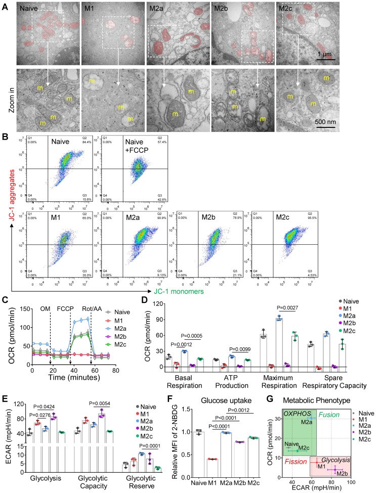
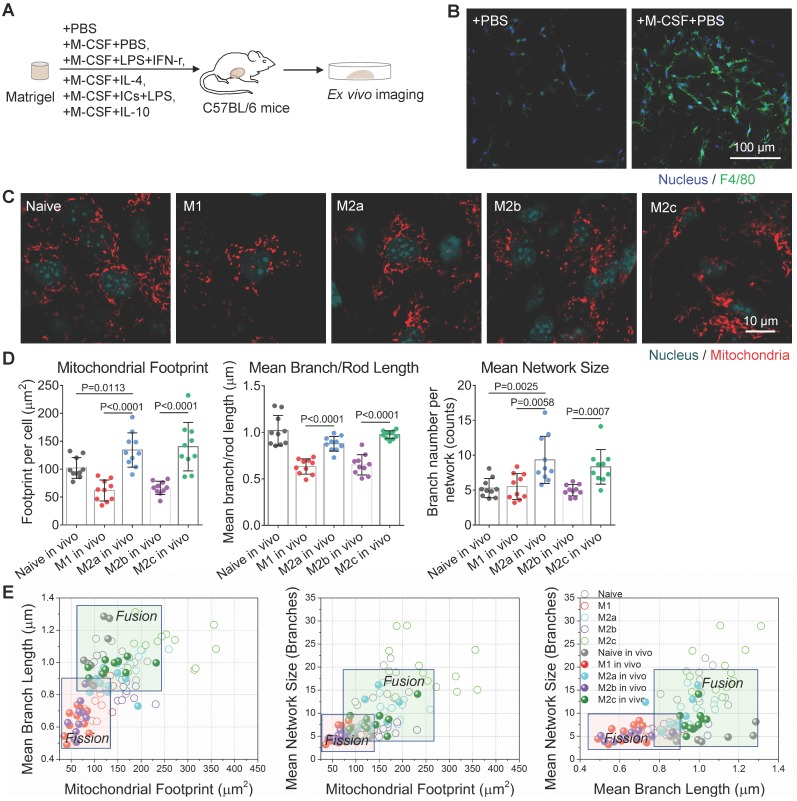
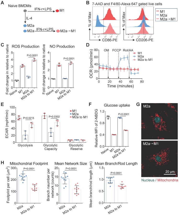
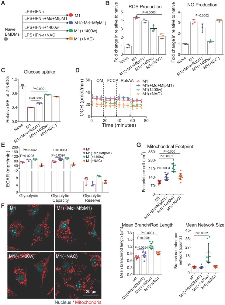
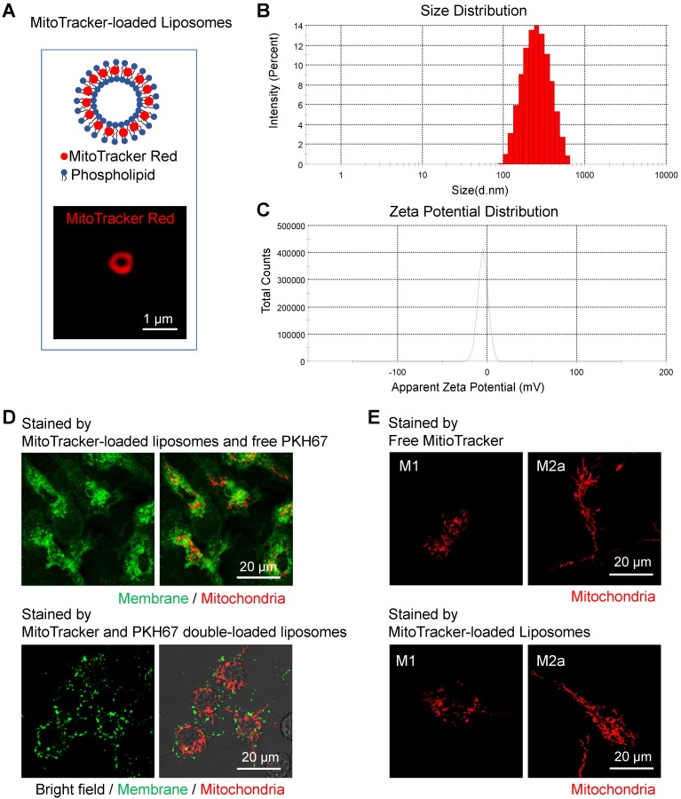
Highly plastic macrophages are pivotal players in the body's homeostasis and pathogenesis. Grasping the molecular or cellular factors that drive and support the macrophage activation will help to develop diagnostics and manipulate their functions in these contexts. However, the lack of characterization methods to reveal the dynamic activation of macrophages impedes these studies in various disease contexts. : Here, bone marrow-derived macrophages (BMDMs) and Matrigel plug were used to evaluate how mitochondria dynamics supports cellular activation and functions. We conducted macrophage repolarization to track mitochondria dynamics during the shift of activation status. For diagnosis, a novel MitoTracker-loaded liposome was first developed to label macrophage mitochondria in mice before/after inflammatory stimulation. : Based on the typical activation of BMDMs, we found glycolysis based macrophages have punctate and discrete mitochondria, while OXPHOS active macrophages have elongated and interconnected mitochondria. M1, M2a, M2b, and M2c activated BMDMs showed clustered and differentiable features in mitochondrial morphology. These features also hold for Matrigel plug-recruited macrophages in mice. Furthermore, with the interventions on M2a macrophages , we demonstrated that mitochondria morphology could be a metabolic index to evaluate macrophage activation status under drug manipulation. Using the MitoTracker-loaded liposomes, we further achieved subcellular imaging of macrophage mitochondria . Their organization dynamics revealed the dynamic change from anti-inflammatory macrophages to inflammatory ones under the lipopolysaccharide (LPS) challenge. : These results reveal that subcellular imaging of mitochondria organization can characterize the activation status of macrophage and at a single-cell level, which is critical for the studies of noninvasive diagnosis and therapeutic drug monitoring.
高塑性巨噬细胞是机体动态平衡和发病机制的关键参与者。了解驱动和支持巨噬细胞激活的分子或细胞因素将有助于开发诊断方法,并在这些情况下操纵其功能。然而,缺乏能够揭示巨噬细胞动态激活的特征方法,阻碍了在各种疾病情况下的这些研究。:在这里,骨髓来源的巨噬细胞(BMDM)和 Matrigel 塞被用来评估线粒体动力学如何支持细胞激活和功能。我们进行了巨噬细胞再极化,以跟踪在激活状态转变过程中线粒体动力学的变化。用于诊断,首先开发了一种新型 MitoTracker 加载的脂质体,以在炎症刺激前后标记小鼠中的巨噬细胞线粒体。:基于 BMDM 的典型激活,我们发现糖酵解依赖的巨噬细胞具有点状和离散的线粒体,而氧化磷酸化(OXPHOS)活跃的巨噬细胞具有伸长和相互连接的线粒体。M1、M2a、M2b 和 M2c 激活的 BMDM 在线粒体形态上表现出聚类和可区分的特征。这些特征在小鼠的 Matrigel 塞募集的巨噬细胞中也存在。此外,通过对 M2a 巨噬细胞的干预,我们证明了线粒体形态可以作为一种代谢指标,用于评估药物干预下巨噬细胞的激活状态。使用 MitoTracker 加载的脂质体,我们进一步实现了巨噬细胞线粒体的亚细胞成像。它们的组织动力学揭示了在脂多糖(LPS)挑战下,从抗炎性巨噬细胞到炎症性巨噬细胞的动态变化。:这些结果表明,亚细胞成像的线粒体组织可以表征巨噬细胞的激活状态,并在单细胞水平上进行,这对于非侵入性诊断和治疗药物监测的研究至关重要。