Unit of Nuclear Medicine, Department of Medical, Surgical and Experimental Sciences, University of Sassari, Viale San Pietro 8, 07100, Sassari, Italy.
BMC Cancer. 2020 Mar 20;20(1):239. doi: 10.1186/s12885-020-06744-1.
The identification of neck lymph node (LN) metastases represents a very important issue in the management of patients with differentiated thyroid carcinoma (DTC). To this purpose, in the present study, we used 131I-SPECT/CT as a diagnostic imaging procedure.
A consecutive series of 224 DTC patients with ascertained neck radioiodine-avid foci at I-SPECT/CT during long-term follow-up was evaluated. All patients had already undergone total thyroidectomy and radioiodine therapy and had been classified as follows: 62 at high risk (H), 64 at low risk (L) and 98 at very low risk (VL). I-Whole body scan (WBS) followed by SPECT/CT was performed in all cases.
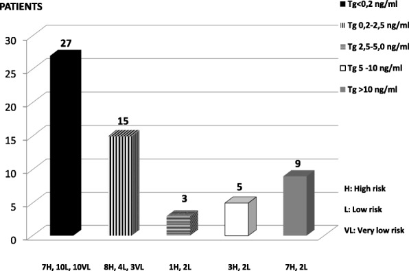
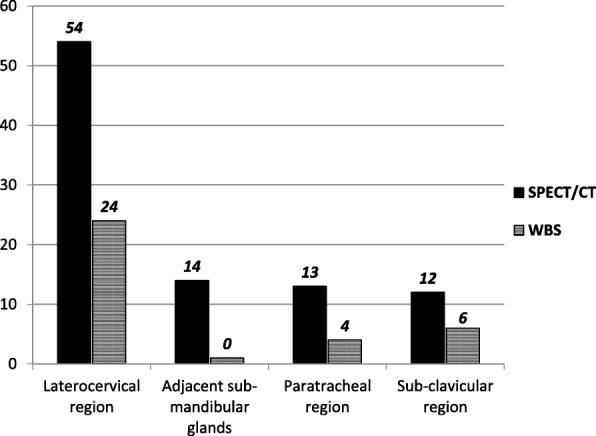
In the 224 patients, 449 neck iodine avid foci were ascertained at SPECT/CT, while 322 were evidenced at WBS in 165/224 patients. WBS classified as residues 263/322 foci and as unclear 59/322 foci; among the former foci SPECT/CT correctly characterized 8 LN metastases and 3 physiologic uptakes and among the latter, it pinpointed 26 LN metastases, 18 residues, and 15 physiologic uptakes. SPECT/CT also classified 127 foci occult at WBS as 59 LN metastases and 68 residues. Globally, SPECT/CT identified 93 LN metastases in 59 patients (26 H, 20 L, 13 VL), while WBS evidenced 34 in 25 cases. All 13 VL patients, T1aN0M0, 5 of whom with LN near sub-mandibular glands, had thyroglobulin undetectable or < 2.5 ng/ml. Globally, SPECT/CT obtained an incremental value than WBS in 45.5% of patients, a more correct patient classification changing therapeutic approach in 30.3% of cases and identified WBS false-positive findings in 8% of cases.
I-SPECT/CT proved to correctly detect and characterize neck LN metastases in DTC patients in long-term follow-up, improving the performance of planar WBS. SPECT/CT routine use is thus suggested; its role is particularly relevant in patients with WBS inconclusive, VL, T1aN0M0 and with undetectable or very low thyroglobulin levels.
在分化型甲状腺癌(DTC)患者的管理中,颈部淋巴结(LN)转移的识别是一个非常重要的问题。为此,在本研究中,我们使用 131I-SPECT/CT 作为诊断成像程序。
连续评估了 224 例在长期随访期间 I-SPECT/CT 证实颈部放射性碘摄取灶的 DTC 患者。所有患者均已接受全甲状腺切除术和放射性碘治疗,并分为以下几类:62 例高危(H),64 例低危(L)和 98 例极低危(VL)。所有患者均进行 I-全身扫描(WBS),然后进行 SPECT/CT。
在 224 例患者中,SPECT/CT 证实 449 个颈部碘摄取灶,而在 165/224 例患者中,WBS 证实 322 个碘摄取灶。WBS 分类为残留 263/322 个病灶和未明确 59/322 个病灶;前者 SPECT/CT 正确诊断出 8 个 LN 转移和 3 个生理性摄取灶,后者 SPECT/CT 准确识别出 26 个 LN 转移,18 个残留灶和 15 个生理性摄取灶。SPECT/CT 还将 WBS 中 127 个隐匿性病灶分类为 59 个 LN 转移和 68 个残留灶。总体而言,SPECT/CT 在 59 例患者(26 例 H,20 例 L,13 例 VL)中识别出 93 个 LN 转移,而 WBS 在 25 例患者中识别出 34 个。所有 13 例 VL 患者,T1aN0M0,其中 5 例 LN 位于颌下腺附近,甲状腺球蛋白均不可检测或<2.5ng/ml。总体而言,SPECT/CT 在 45.5%的患者中具有比 WBS 更高的增值价值,在 30.3%的病例中更正确地分类患者,改变了治疗方法,并在 8%的病例中识别出 WBS 假阳性发现。
I-SPECT/CT 可在 DTC 患者的长期随访中正确检测和特征化颈部 LN 转移,从而提高平面 WBS 的性能。因此,建议常规使用 SPECT/CT;其作用在 WBS 不确定、VL、T1aN0M0 和甲状腺球蛋白不可检测或极低的患者中尤其重要。