Department of Neurosurgery, Emory University School of Medicine, Atlanta, GA, USA.
Department of Biomedical Engineering, Georgia Institute of Technology, Atlanta, GA, USA.
Sci Rep. 2020 Mar 24;10(1):5291. doi: 10.1038/s41598-020-62167-9.
Prior studies have applied driver mutations targeting the RTK/RAS/PI3K and p53 pathways to induce the formation of high-grade gliomas in rodent models. In the present study, we report the production of a high-grade spinal cord glioma model in pigs using lentiviral gene transfer.
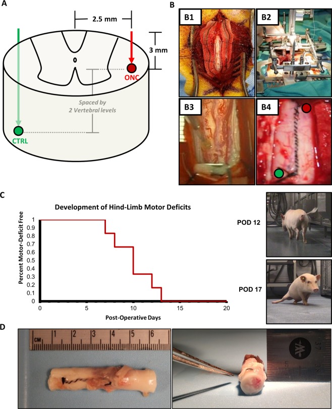
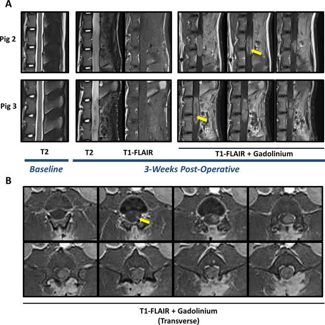
Six Gottingen Minipigs received thoracolumbar (T14-L1) lateral white matter injections of a combination of lentiviral vectors, expressing platelet-derived growth factor beta (PDGF-B), constitutive HRAS, and shRNA-p53 respectively. All animals received injection of control vectors into the contralateral cord. Animals underwent baseline and endpoint magnetic resonance imaging (MRI) and were evaluated daily for clinical deficits. Hematoxylin and eosin (H&E) and immunohistochemical analysis was conducted. Data are presented using descriptive statistics including relative frequencies, mean, standard deviation, and range.
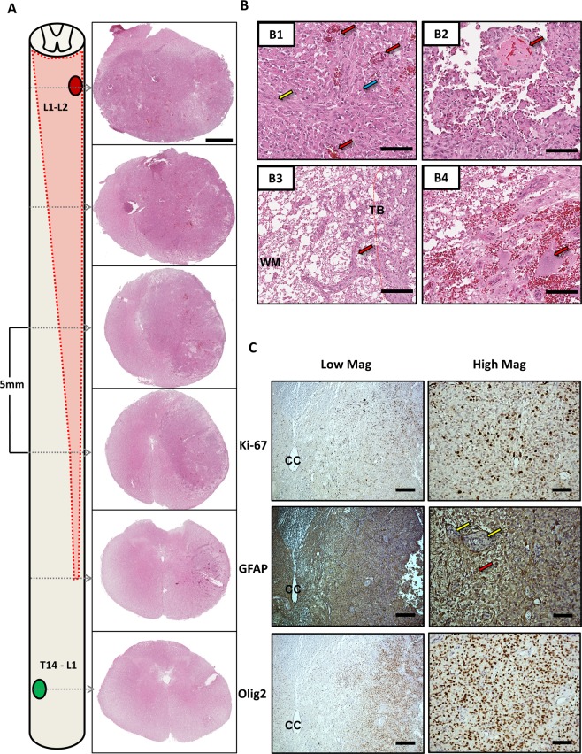
100% of animals (n = 6/6) developed clinical motor deficits ipsilateral to the oncogenic lentiviral injections by a three-week endpoint. MRI scans at endpoint demonstrated contrast enhancing mass lesions at the site of oncogenic lentiviral injection and not at the site of control injections. Immunohistochemistry demonstrated positive staining for GFAP, Olig2, and a high Ki-67 proliferative index. Histopathologic features demonstrate consistent and reproducible growth of a high-grade glioma in all animals.
Lentiviral gene transfer represents a feasible pathway to glioma modeling in higher order species. The present model is the first lentiviral vector induced pig model of high-grade spinal cord glioma and may potentially be used in preclinical therapeutic development programs.
先前的研究已经应用针对 RTK/RAS/PI3K 和 p53 通路的驱动基因突变来诱导啮齿动物模型中高级别脑胶质瘤的形成。在本研究中,我们报告了使用慢病毒基因转移在猪中产生高级别脊髓神经胶质瘤模型。
六只哥廷根小型猪接受胸腰段(T14-L1)侧白质的慢病毒载体组合注射,分别表达血小板衍生生长因子β(PDGF-B)、组成型 HRAS 和 shRNA-p53。所有动物均在对侧脊髓接受对照载体注射。动物进行基线和终点磁共振成像(MRI)检查,并每天进行临床缺陷评估。进行苏木精和伊红(H&E)和免疫组织化学分析。数据使用描述性统计,包括相对频率、平均值、标准差和范围。
100%的动物(n=6/6)在三周末点出现了致癌性慢病毒注射侧的临床运动缺陷。终点 MRI 扫描显示在致癌性慢病毒注射部位有对比增强的肿块病变,而在对照注射部位没有。免疫组织化学显示 GFAP、Olig2 和高 Ki-67 增殖指数阳性染色。组织病理学特征显示所有动物均有一致且可重复的高级别胶质瘤生长。
慢病毒基因转移代表了在高级别物种中进行胶质瘤建模的可行途径。本模型是第一个慢病毒载体诱导的猪高级别脊髓神经胶质瘤模型,可能可用于临床前治疗开发计划。