• 文献检索
  • 文档翻译
  • 深度研究
  • 学术资讯
  • Suppr Zotero 插件Zotero 插件
  • 邀请有礼
  • 套餐&价格
  • 历史记录
应用&插件
Suppr Zotero 插件Zotero 插件浏览器插件Mac 客户端Windows 客户端微信小程序
定价
高级版会员购买积分包购买API积分包
服务
文献检索文档翻译深度研究API 文档MCP 服务
关于我们
关于 Suppr公司介绍联系我们用户协议隐私条款
关注我们

Suppr 超能文献

核心技术专利:CN118964589B侵权必究
粤ICP备2023148730 号-1Suppr @ 2026

文献检索

告别复杂PubMed语法,用中文像聊天一样搜索,搜遍4000万医学文献。AI智能推荐,让科研检索更轻松。

立即免费搜索

文件翻译

保留排版,准确专业,支持PDF/Word/PPT等文件格式,支持 12+语言互译。

免费翻译文档

深度研究

AI帮你快速写综述,25分钟生成高质量综述,智能提取关键信息,辅助科研写作。

立即免费体验

肺动脉充盈缺损,一项我们应该了解的影像学表现。

The filling defect of pulmonary artery, an imaging finding what we should know.

作者信息

Liu Min, Tao Xin Cao, Zhai Zhenguo, Ma Zhanhong, Zhu Li, Luo Jie

机构信息

Department of Radiology, China-Japan Friendship Hospital, Beijing, China.

Department of Pulmonary and Critical Care Medicine, China-Japan Friendship Hospital, Beijing, China.

出版信息

Pulm Circ. 2020 Mar 6;10(1):2045894020910687. doi: 10.1177/2045894020910687. eCollection 2020 Jan-Mar.

DOI:10.1177/2045894020910687
PMID:32215201
原文链接:https://pmc.ncbi.nlm.nih.gov/articles/PMC7065294/
Abstract

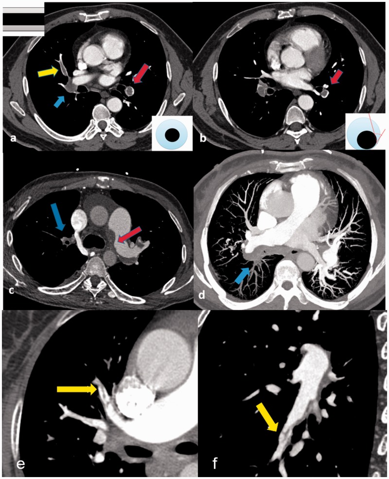
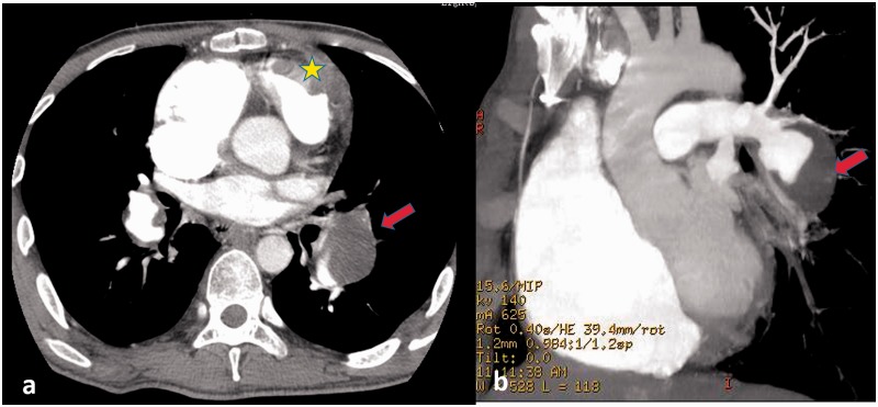
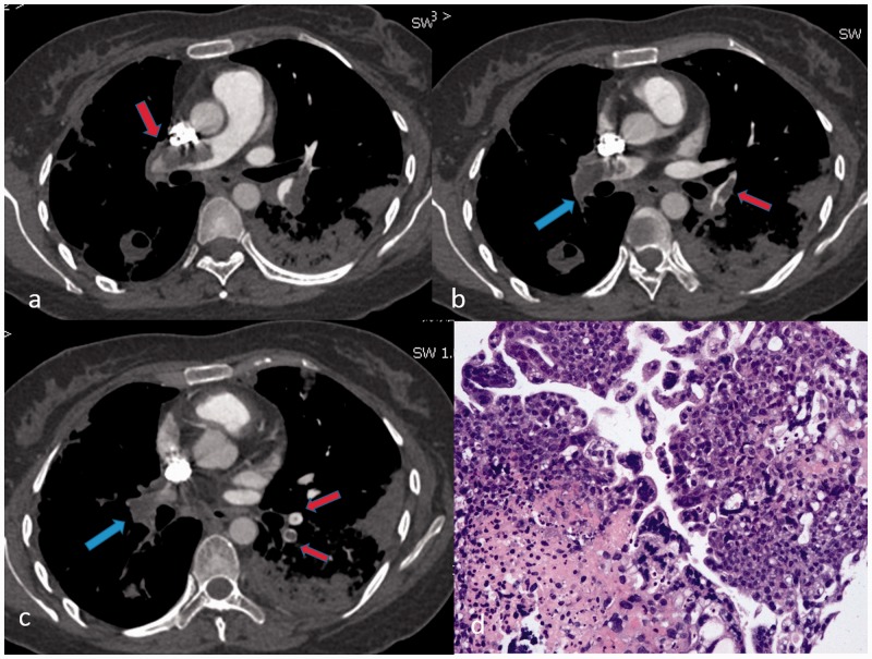
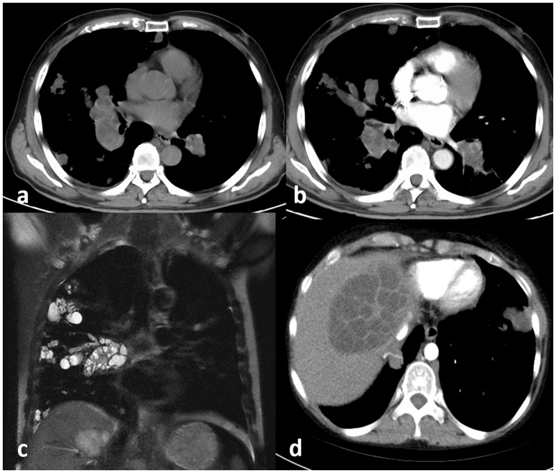
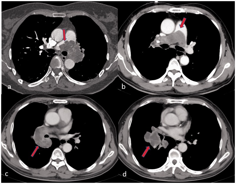
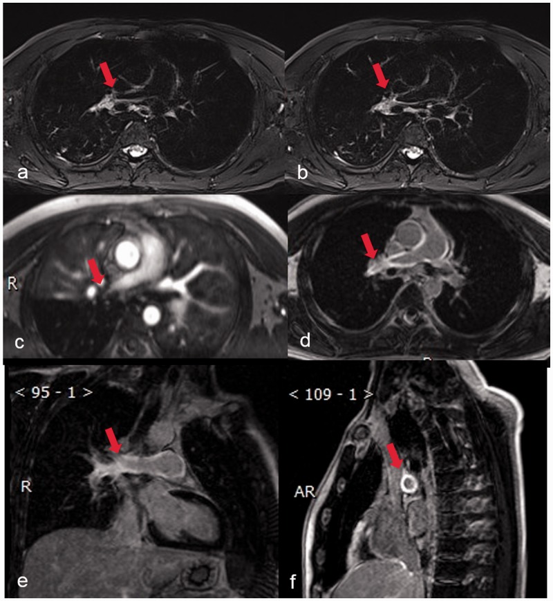
The most common cause of pulmonary artery filling defects on computed tomography pulmonary angiography or magnetic resonance imaging is pulmonary thromboembolism, but not infrequently, the presentation of this finding lacks specificity. Given that the morbidity and mortality associated with pulmonary thromboembolism is high, proper diagnosis of the condition is essential. Unusual or more rarely encountered etiologies must be considered when clinical manifestations and imaging findings are inconsistent. With this review, our purpose is to describe possible causes of pulmonary arterial filling defects. We aim to provide clinicians with a comprehensive list of differential diagnoses to facilitate a measured approach to the assessment of pulmonary arterial filling defects on computed tomography pulmonary angiography or magnetic resonance imaging.

摘要

计算机断层扫描肺动脉造影或磁共振成像上肺动脉充盈缺损的最常见原因是肺血栓栓塞,但这种表现往往缺乏特异性。鉴于肺血栓栓塞相关的发病率和死亡率很高,正确诊断该疾病至关重要。当临床表现和影像学表现不一致时,必须考虑不常见或更罕见的病因。通过本综述,我们的目的是描述肺动脉充盈缺损的可能原因。我们旨在为临床医生提供一份全面的鉴别诊断清单,以便在计算机断层扫描肺动脉造影或磁共振成像上对肺动脉充盈缺损进行评估时采取审慎的方法。

https://cdn.ncbi.nlm.nih.gov/pmc/blobs/40fb/7065294/4f89fa95b531/10.1177_2045894020910687-fig6.jpg
https://cdn.ncbi.nlm.nih.gov/pmc/blobs/40fb/7065294/4748517de016/10.1177_2045894020910687-fig1.jpg
https://cdn.ncbi.nlm.nih.gov/pmc/blobs/40fb/7065294/5d77579cf53d/10.1177_2045894020910687-fig2.jpg
https://cdn.ncbi.nlm.nih.gov/pmc/blobs/40fb/7065294/32810a82b529/10.1177_2045894020910687-fig3.jpg
https://cdn.ncbi.nlm.nih.gov/pmc/blobs/40fb/7065294/3d0021a91581/10.1177_2045894020910687-fig4.jpg
https://cdn.ncbi.nlm.nih.gov/pmc/blobs/40fb/7065294/0fbf2f894d3f/10.1177_2045894020910687-fig5.jpg
https://cdn.ncbi.nlm.nih.gov/pmc/blobs/40fb/7065294/4f89fa95b531/10.1177_2045894020910687-fig6.jpg

相似文献

1
The filling defect of pulmonary artery, an imaging finding what we should know.肺动脉充盈缺损,一项我们应该了解的影像学表现。
Pulm Circ. 2020 Mar 6;10(1):2045894020910687. doi: 10.1177/2045894020910687. eCollection 2020 Jan-Mar.
2
Systemic Artery-to-Pulmonary Artery Fistula Mimics Pulmonary Embolus.体动脉至肺动脉瘘酷似肺栓塞。
J Clin Imaging Sci. 2019 Sep 20;9:41. doi: 10.25259/JCIS_54_2019. eCollection 2019.
3
Unusual Pulmonary Arterial Filling Defect caused by Systemic to Pulmonary Shunt in the Setting of Chronic Lung Disease Demonstrated by Dynamic 4D CTA.动态4D CTA显示慢性肺病背景下由体肺分流引起的罕见肺动脉充盈缺损
J Radiol Case Rep. 2015 Nov 30;9(11):17-23. doi: 10.3941/jrcr.v9i11.2480. eCollection 2015 Nov.
4
Differential Diagnosis of Pulmonary Artery Sarcoma and Central Chronic Pulmonary Thromboembolism Using CT and MR Images.利用CT和MR图像对肺动脉肉瘤与中央型慢性肺血栓栓塞症进行鉴别诊断
Heart Lung Circ. 2018 Jul;27(7):819-827. doi: 10.1016/j.hlc.2017.06.716. Epub 2017 Jul 1.
5
Diagnosis of pulmonary arterial hypertension and pulmonary embolism with magnetic resonance angiography.
Chest. 2001 Nov;120(5):1556-61. doi: 10.1378/chest.120.5.1556.
6
[The clinical and CT two-phase imaging features of bronchial-pulmonary arterial fistula].[支气管-肺动脉瘘的临床及CT双期影像学特征]
Zhonghua Jie He He Hu Xi Za Zhi. 2014 Sep;37(9):687-93.
7
A new perspective for pulmonary thromboembolism radiology: 'Pulmonary vein sign'.肺血栓栓塞症放射学的新视角:“肺静脉征”
Clin Respir J. 2018 Dec;12(12):2653-2658. doi: 10.1111/crj.12971. Epub 2018 Nov 13.
8
[Helical CT as preferred imaging modality in the diagnosis of pulmonary embolism].
Radiol Med. 2001 Nov-Dec;102(5-6):320-4.
9
Delayed systemic arterial-pulmonary arterial shunt simulating pulmonary embolism: An unusual case.模拟肺栓塞的延迟性体动脉-肺动脉分流:一例罕见病例。
Radiol Case Rep. 2024 Aug 27;19(11):5271-5275. doi: 10.1016/j.radcr.2024.07.177. eCollection 2024 Nov.
10
Pulmonary Arterial Hypertension With Abnormal V/Q Single-Photon Emission Computed Tomography.肺动脉高压伴异常通气/血流比单光子发射计算机断层扫描。
JACC Cardiovasc Imaging. 2018 Oct;11(10):1487-1493. doi: 10.1016/j.jcmg.2017.07.026. Epub 2017 Oct 18.

引用本文的文献

1
CT features and risk factors of pulmonary cement embolism after vertebroplasty or kyphoplasty in patients with vertebral compression fracture: a retrospective cohort study.椎体压缩骨折患者椎体成形术或后凸成形术后肺水泥栓塞的CT特征及危险因素:一项回顾性队列研究
Quant Imaging Med Surg. 2023 Apr 1;13(4):2397-2407. doi: 10.21037/qims-22-569. Epub 2023 Feb 27.
2
Improving the imaging diagnostic strategy for pulmonary artery masses based on F-FDG PET/CT integrated with CTPA.基于F-FDG PET/CT与CTPA相结合改善肺动脉肿块的影像诊断策略。
Eur J Nucl Med Mol Imaging. 2022 Oct;49(12):4109-4121. doi: 10.1007/s00259-022-05851-4. Epub 2022 Jun 23.
3

本文引用的文献

1
Pulmonary vascular involvement of IgG4-related disease: Case series with a PRISMA-compliant systemic review.IgG4相关性疾病的肺血管受累:符合PRISMA标准的系统评价病例系列
Medicine (Baltimore). 2019 Feb;98(6):e14437. doi: 10.1097/MD.0000000000014437.
2
Intraluminal Arterial Filling Defects Misdiagnosed as Pulmonary Emboli: What Else Could They Be?腔内动脉充盈缺损被误诊为肺栓塞:还可能是什么?
Clin Chest Med. 2018 Sep;39(3):505-513. doi: 10.1016/j.ccm.2018.04.004.
3
Primary Pulmonary Artery Lipoma Mimicking Pulmonary Thromboembolism.
Pulmonary artery intimal sarcoma mimicking pulmonary thromboembolism: A case report.
肺动脉内膜肉瘤酷似肺血栓栓塞症:1 例报告。
Medicine (Baltimore). 2021 Feb 12;100(6):e24699. doi: 10.1097/MD.0000000000024699.
4
Misdiagnosis of Pulmonary embolism in a Fontan's patient: When standard protocol CT Pulmonary angiogram is inadequate.法洛四联症患者的肺栓塞误诊:当标准方案CT肺动脉造影不充分时。
Radiol Case Rep. 2020 Sep 10;15(11):2262-2265. doi: 10.1016/j.radcr.2020.08.041. eCollection 2020 Nov.
5
To reperfuse or not to reperfuse: a case report of Wellens' syndrome with suspected COVID-19 infection.是否进行再灌注治疗:一例疑似新型冠状病毒肺炎感染的Wellens综合征病例报告
Egypt Heart J. 2020 Sep 9;72:58. doi: 10.1186/s43044-020-00094-w. eCollection 2020 Dec.
Am J Respir Crit Care Med. 2018 Nov 15;198(10):e111-e113. doi: 10.1164/rccm.201706-1151IM.
4
Pulmonary Endarterectomy For Large Vessel Pulmonary Arteritis Mimicking Chronic Thromboembolic Disease.针对酷似慢性血栓栓塞性疾病的大血管性肺动脉炎行肺动脉内膜剥脱术
Arch Rheumatol. 2015 Nov 2;31(1):98-101. doi: 10.5606/ArchRheumatol.2016.5674. eCollection 2016 Mar.
5
Chronic Thromboembolic Pulmonary Hypertension: Evolving Therapeutic Approaches for Operable and Inoperable Disease.慢性血栓栓塞性肺动脉高压:可手术和不可手术疾病的治疗方法的进展。
J Am Coll Cardiol. 2018 May 29;71(21):2468-2486. doi: 10.1016/j.jacc.2018.04.009.
6
IgG4-related disease of pulmonary artery causing pulmonary hypertension.IgG4相关性肺动脉疾病导致肺动脉高压。
Medicine (Baltimore). 2018 May;97(20):e10698. doi: 10.1097/MD.0000000000010698.
7
F-18 FDG PET/CT in pulmonary artery sarcoma: clinical vignette.18F-FDG PET/CT在肺动脉肉瘤中的应用:临床病例
Nucl Med Rev Cent East Eur. 2018;21(1):48-49. doi: 10.5603/NMR.a2018.0011.
8
Evaluation of takayasu arteritis with delayed contrast-enhanced MR imaging by a free-breathing 3D IR turbo FLASH.自由呼吸三维反转恢复快速低角度激发磁共振成像延迟增强对高安动脉炎的评估
Medicine (Baltimore). 2017 Dec;96(51):e9284. doi: 10.1097/MD.0000000000009284.
9
CT Features of Vasculitides Based on the 2012 International Chapel Hill Consensus Conference Revised Classification.基于2012年国际教堂山共识会议修订分类的血管炎的CT特征
Korean J Radiol. 2017 Sep-Oct;18(5):786-798. doi: 10.3348/kjr.2017.18.5.786. Epub 2017 Jul 17.
10
A Rare Cause of Pulmonary Embolism: Hydatid Cyst.肺栓塞的罕见病因:包虫囊肿
Am J Respir Crit Care Med. 2017 Aug 1;196(3):386-387. doi: 10.1164/rccm.201612-2459IM.