Jiang Hao, Zhang Rongjun, Jiang Huijie, Zhang Mingyu, Guo Wei, Zhang Jifeng, Zhou Xinglu, Pan Wenbin, Zhao Sheng, Li Ping
Department of Radiology, The Second Affiliated Hospital of Harbin Medical University, Harbin, China.
Jiangsu Institute of Nuclear Medicine, Wuxi, China.
J Cancer. 2020 Feb 25;11(10):2864-2873. doi: 10.7150/jca.38689. eCollection 2020.
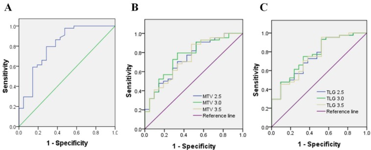
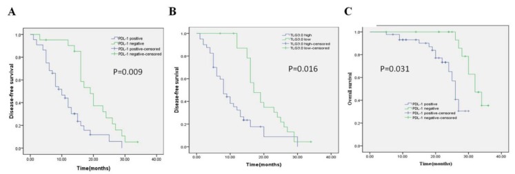
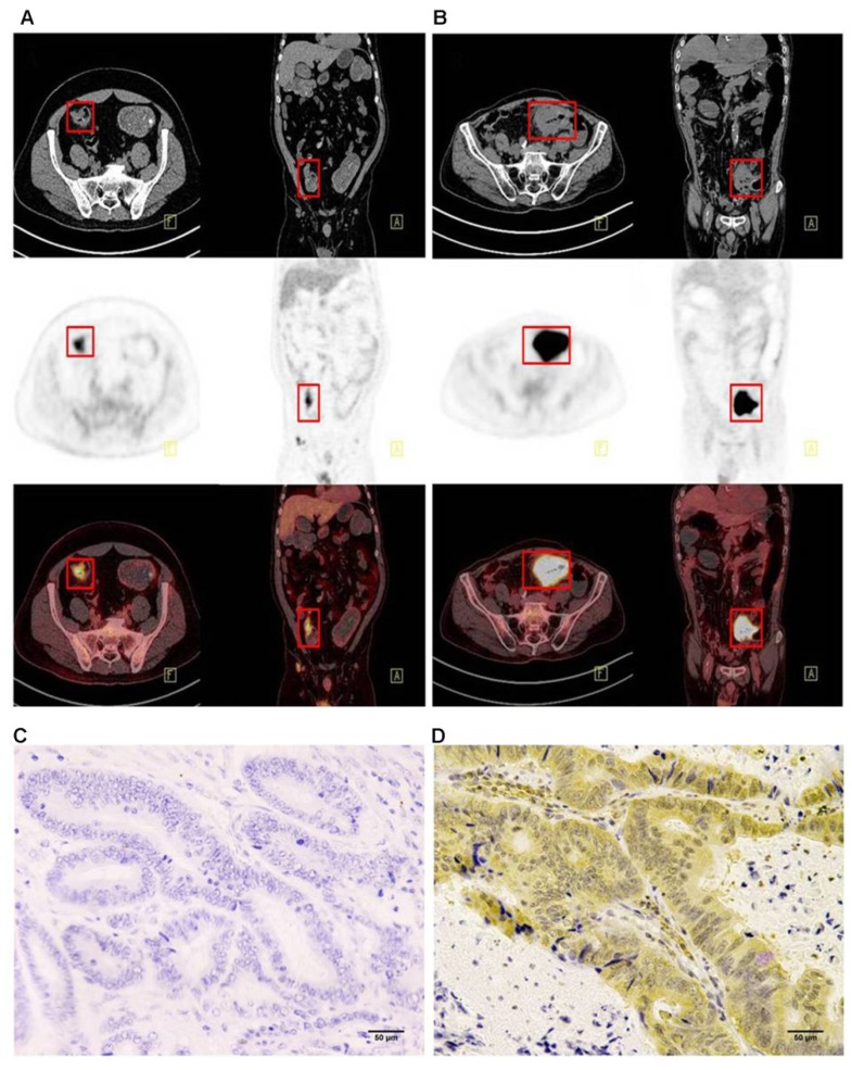
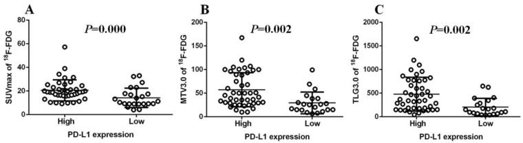
It has been rarely reported whether F-fluorodeoxyglucose (F-FDG) uptake in colorectal cancer cells is associated with the expression of PD-L1. We performed a clinical pathology study to evaluate PD-L1 expression in patients undergoing surgical resection of colorectal cancer with preoperative F-FDG PET/CT imaging, with the aim of predicting the response of CRC patients to immune checkpoint inhibitors. : A retrospective analysis of patients with CRC who underwent FDG-PET imaging before surgery was performed to measure the parameters of FDG-PET imaging: the maximum standardized uptake value (SUVmax), the metabolic tumor volume (MTV), and the total lesion glycolysis (TLG) were evaluated to determine whether each parameter was associated with clinical pathology. Tumor specimens were subjected to PD-L1 staining by immunohistochemistry. Analysis of whether there is a correlation between PD-L1 expression and F-FDG uptake parameters in CRC. : PD-L1 expression level was significantly correlated with SUVmax, MTV3.0 and TLG3.0. Multivariate analysis showed that PD-L1 and TLG3.0 were independent predictors of poor DFS in patients with CRC (0.009; 0.016), PD-L1 expression is closely related to the patient's lesion (TLG3.0) (<0.01). : The results of this study indicate that there was a significant correlation between PD-L1 expression and TLG3.0 which suggested that FDG-PET could serve as a noninvasive tool to assess the tumor microenvironment and as a predictor of PD-L1 inhibitor activity to determine the optimal therapeutic strategy for CRC. High PD-L1 expression levels and high TLG3.0 are independent risk factors for DFS differences in CRC patients.
结直肠癌细胞中氟脱氧葡萄糖(F-FDG)摄取是否与程序性死亡受体配体1(PD-L1)的表达相关鲜有报道。我们进行了一项临床病理研究,以评估术前接受F-FDG PET/CT成像的结直肠癌手术切除患者的PD-L1表达,目的是预测结直肠癌患者对免疫检查点抑制剂的反应。:对术前接受FDG-PET成像的结直肠癌患者进行回顾性分析,以测量FDG-PET成像参数:评估最大标准化摄取值(SUVmax)、代谢肿瘤体积(MTV)和总病变糖酵解(TLG),以确定每个参数是否与临床病理相关。肿瘤标本通过免疫组织化学进行PD-L1染色。分析结直肠癌中PD-L1表达与F-FDG摄取参数之间是否存在相关性。:PD-L1表达水平与SUVmax、MTV3.0和TLG3.0显著相关。多因素分析表明,PD-L1和TLG3.0是结直肠癌患者无病生存期差的独立预测因素(0.009;0.016),PD-L1表达与患者病变(TLG3.0)密切相关(<0.01)。:本研究结果表明,PD-L1表达与TLG3.0之间存在显著相关性,这表明FDG-PET可作为评估肿瘤微环境的非侵入性工具,以及作为PD-L1抑制剂活性的预测指标,以确定结直肠癌的最佳治疗策略。高PD-L1表达水平和高TLG3.0是结直肠癌患者无病生存期差异的独立危险因素。