• 文献检索
  • 文档翻译
  • 深度研究
  • 学术资讯
  • Suppr Zotero 插件Zotero 插件
  • 邀请有礼
  • 套餐&价格
  • 历史记录
应用&插件
Suppr Zotero 插件Zotero 插件浏览器插件Mac 客户端Windows 客户端微信小程序
定价
高级版会员购买积分包购买API积分包
服务
文献检索文档翻译深度研究API 文档MCP 服务
关于我们
关于 Suppr公司介绍联系我们用户协议隐私条款
关注我们

Suppr 超能文献

核心技术专利:CN118964589B侵权必究
粤ICP备2023148730 号-1Suppr @ 2026

文献检索

告别复杂PubMed语法,用中文像聊天一样搜索,搜遍4000万医学文献。AI智能推荐,让科研检索更轻松。

立即免费搜索

文件翻译

保留排版,准确专业,支持PDF/Word/PPT等文件格式,支持 12+语言互译。

免费翻译文档

深度研究

AI帮你快速写综述,25分钟生成高质量综述,智能提取关键信息,辅助科研写作。

立即免费体验

定量磁化率成像和血管壁成像作为检测哨兵头痛中微出血的筛查工具。

Quantitative Susceptibility Mapping and Vessel Wall Imaging as Screening Tools to Detect Microbleed in Sentinel Headache.

作者信息

Ishii Daizo, Nakagawa Daichi, Zanaty Mario, Roa Jorge A, Al Kasab Sami, Shaban Amir, Hudson Joseph S, Osorno-Cruz Carlos, Byer Stefano, Allan Lauren, Torner James C, Awad Issam A, Carroll Timothy J, Samaniego Edgar A, Hasan David M

机构信息

Department of Neurosurgery, University of Iowa Hospitals and Clinics, Iowa City, IA 52242, USA.

Department of Neurosurgery, University of Tokyo, Tokyo 113 8654, Japan.

出版信息

J Clin Med. 2020 Apr 1;9(4):979. doi: 10.3390/jcm9040979.

DOI:10.3390/jcm9040979
PMID:32244737
原文链接:https://pmc.ncbi.nlm.nih.gov/articles/PMC7230854/
Abstract

BACKGROUND

MR-quantitative susceptibility mapping (QSM) can identify microbleeds (MBs) in intracranial aneurysm (IA) wall associated with sentinel headache (SH) preceding subarachnoid hemorrhage. However, its use is limited, due to associated skull base bonny and air artifact. MR-vessel wall imaging (VWI) is not limited by such artifact and therefore could be an alternative to QSM. The purpose of this study was to investigate the correlation between QSM and VWI in detecting MBs and to help develop a diagnostic strategy for SH.

METHODS

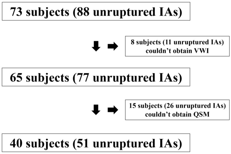
We performed a prospective study of subjects with one or more unruptured IAs in our hospital. All subjects underwent evaluation using 3T-MRI for MR angiography (MRA), QSM, and pre- and post-contrast VWI of the IAs. Presence/absence of MBs detected by QSM was correlated with aneurysm wall enhancement (AWE) on VWI.

RESULTS

A total of 40 subjects harboring 51 unruptured IAs were enrolled in the study. MBs evident on the QSM sequence was detected in 12 (23.5%) IAs of 11 subjects. All these subjects had a history of severe headache suggestive of SH. AWE was detected in 22 (43.1%) IAs. Using positive QSM as a surrogate for MBs, the sensitivity, specificity, positive predictive value, and negative predictive value of AWE on VWI for detecting MBs were 91.7%, 71.8%, 50%, and 96.6%, respectively.

CONCLUSIONS

Positive QSM findings strongly suggested the presence of MBs with SH, whereas, the lack of AWE on VWI can rule it out with a probability of 96.6%. If proven in a larger cohort, combining QSM and VWI could be an adjunctive tool to help diagnose SH, especially in cases with negative or non-diagnostic CT and lumbar puncture.

摘要

背景

磁共振定量磁化率成像(QSM)能够识别颅内动脉瘤(IA)壁内与蛛网膜下腔出血前的哨兵头痛(SH)相关的微出血(MBs)。然而,由于存在颅底骨质和空气伪影,其应用受到限制。磁共振血管壁成像(VWI)不受此类伪影的限制,因此可能是QSM的替代方法。本研究的目的是探讨QSM与VWI在检测MBs方面的相关性,并帮助制定SH的诊断策略。

方法

我们对我院有一个或多个未破裂IA的受试者进行了一项前瞻性研究。所有受试者均接受3T-MRI检查,以进行磁共振血管造影(MRA)、QSM以及IA的对比剂增强前后VWI检查。QSM检测到的MBs的有无与VWI上的动脉瘤壁强化(AWE)相关。

结果

本研究共纳入了40例患有51个未破裂IA的受试者。在11例受试者的12个(23.5%)IA中检测到QSM序列上明显的MBs。所有这些受试者都有提示SH的严重头痛病史。在22个(43.1%)IA中检测到AWE。以阳性QSM作为MBs的替代指标,VWI上AWE检测MBs的敏感性、特异性、阳性预测值和阴性预测值分别为91.7%、71.8%、50%和96.6%。

结论

QSM阳性结果强烈提示存在伴有SH的MBs,而VWI上缺乏AWE则可在96.6%的概率上排除。如果在更大的队列中得到证实,联合使用QSM和VWI可能是一种辅助工具,有助于诊断SH,尤其是在CT和腰椎穿刺结果为阴性或无法诊断的情况下。

https://cdn.ncbi.nlm.nih.gov/pmc/blobs/76f6/7230854/fd7d5b12a300/jcm-09-00979-g003.jpg
https://cdn.ncbi.nlm.nih.gov/pmc/blobs/76f6/7230854/3e3c8c63eb7e/jcm-09-00979-g001.jpg
https://cdn.ncbi.nlm.nih.gov/pmc/blobs/76f6/7230854/2eb2478f877a/jcm-09-00979-g002.jpg
https://cdn.ncbi.nlm.nih.gov/pmc/blobs/76f6/7230854/fd7d5b12a300/jcm-09-00979-g003.jpg
https://cdn.ncbi.nlm.nih.gov/pmc/blobs/76f6/7230854/3e3c8c63eb7e/jcm-09-00979-g001.jpg
https://cdn.ncbi.nlm.nih.gov/pmc/blobs/76f6/7230854/2eb2478f877a/jcm-09-00979-g002.jpg
https://cdn.ncbi.nlm.nih.gov/pmc/blobs/76f6/7230854/fd7d5b12a300/jcm-09-00979-g003.jpg

相似文献

1
Quantitative Susceptibility Mapping and Vessel Wall Imaging as Screening Tools to Detect Microbleed in Sentinel Headache.定量磁化率成像和血管壁成像作为检测哨兵头痛中微出血的筛查工具。
J Clin Med. 2020 Apr 1;9(4):979. doi: 10.3390/jcm9040979.
2
Quantitative Susceptibility Mapping as a Possible Tool to Radiographically Diagnose Sentinel Headache Associated with Intracranial Aneurysm: Case Report.定量磁化率图谱作为影像学诊断颅内动脉瘤相关前驱性头痛的一种可能工具:病例报告
World Neurosurg. 2017 Jul;103:954.e1-954.e4. doi: 10.1016/j.wneu.2017.04.151. Epub 2017 Apr 30.
3
Neutrophil-to-Lymphocyte Ratio Is Associated With Circumferential Wall Enhancement of Unruptured Intracranial Aneurysm.中性粒细胞与淋巴细胞比值与未破裂颅内动脉瘤的环壁强化有关。
Front Neurol. 2022 May 20;13:879882. doi: 10.3389/fneur.2022.879882. eCollection 2022.
4
Vessel wall enhancement of intracranial aneurysms: fact or artifact?颅内动脉瘤的血管壁增强:是事实还是伪影?
Neurosurg Focus. 2019 Jul 1;47(1):E18. doi: 10.3171/2019.4.FOCUS19236.
5
Vessel Wall Imaging Predicts the Presence of Atherosclerotic Lesions in Unruptured Intracranial Aneurysms.血管壁成像可预测未破裂颅内动脉瘤中的动脉粥样硬化病变。
World Neurosurg. 2019 Dec;132:e775-e782. doi: 10.1016/j.wneu.2019.08.019. Epub 2019 Aug 12.
6
Detection of microbleeds associated with sentinel headache using MRI quantitative susceptibility mapping: pilot study.使用MRI定量磁化率成像检测与前驱性头痛相关的微出血:初步研究
J Neurosurg. 2018 May 25;130(4):1391-1397. doi: 10.3171/2018.2.JNS1884. Print 2019 Apr 1.
7
Relationship between aneurysm wall enhancement and conventional risk factors in patients with unruptured intracranial aneurysms: A black-blood MRI study.未破裂颅内动脉瘤患者动脉瘤壁强化与传统危险因素的关系:一项黑血MRI研究
Interv Neuroradiol. 2016 Oct;22(5):501-5. doi: 10.1177/1591019916653252. Epub 2016 Jun 24.
8
Role of magnetic resonance vessel wall imaging in detecting and managing ruptured aneurysms among multiple intracranial aneurysms.磁共振血管壁成像在检测和处理多发性颅内动脉瘤中破裂动脉瘤方面的作用
Surg Neurol Int. 2021 Sep 13;12:460. doi: 10.25259/SNI_618_2021. eCollection 2021.
9
Relationship Between Aneurysm Wall Enhancement in Vessel Wall Magnetic Resonance Imaging and Rupture Risk of Unruptured Intracranial Aneurysms.颅内未破裂动脉瘤瘤壁增强与破裂风险的关系:血管壁磁共振成像研究
Neurosurgery. 2019 Jun 1;84(6):E385-E391. doi: 10.1093/neuros/nyy310.
10
Segmentation of aneurysm wall enhancement in evolving unruptured intracranial aneurysms.颅内未破裂进展性动脉瘤瘤壁增强的分割。
J Neurosurg. 2021 Aug 13;136(2):449-455. doi: 10.3171/2021.2.JNS2114. Print 2022 Feb 1.

引用本文的文献

1
Beyond Size: Advanced MRI Breakthroughs in Predicting Intracranial Aneurysm Rupture Risk.超越大小:预测颅内动脉瘤破裂风险的先进磁共振成像突破
J Clin Med. 2025 May 2;14(9):3158. doi: 10.3390/jcm14093158.
2
Radiomics-Based Predictive Nomogram for Assessing the Risk of Intracranial Aneurysms.基于影像组学的预测列线图用于评估颅内动脉瘤风险
Res Sq. 2024 May 10:rs.3.rs-4350156. doi: 10.21203/rs.3.rs-4350156/v1.
3
Feasibility and intra-and interobserver reproducibility of quantitative susceptibility mapping with radiomic features for intracranial dissecting intramural hematomas and atherosclerotic calcifications.

本文引用的文献

1
Vessel Wall Imaging of Evolving Unruptured Intracranial Aneurysms.颅内未破裂动脉瘤演变的血管壁成像。
Stroke. 2019 Jul;50(7):1891-1894. doi: 10.1161/STROKEAHA.119.025245. Epub 2019 Jun 6.
2
Quantitative Susceptibility Mapping for Carotid Atherosclerotic Plaques: A Pilot Study.颈动脉粥样硬化斑块的定量磁敏感图:一项初步研究。
Magn Reson Med Sci. 2020 May 1;19(2):135-140. doi: 10.2463/mrms.mp.2018-0077. Epub 2019 May 31.
3
Grading meningiomas utilizing multiparametric MRI with inclusion of susceptibility weighted imaging and quantitative susceptibility mapping.
颅内夹层壁内血肿和动脉粥样硬化钙化的定量磁化率映射与放射组学特征的可行性及观察者内和观察者间可重复性。
Sci Rep. 2023 Mar 4;13(1):3651. doi: 10.1038/s41598-023-30745-2.
4
Diagnostic criteria for acute headache attributed to ischemic stroke and for sentinel headache before ischemic stroke.急性缺血性脑卒中相关性头痛和缺血性脑卒中前预警性头痛的诊断标准。
J Headache Pain. 2022 Jan 20;23(1):11. doi: 10.1186/s10194-021-01372-x.
5
Warning Signs in the Era of Unruptured Intracranial Aneurysms: Report on 2 Cases of Fatal Aneurysmal Hemorrhage.未破裂颅内动脉瘤时代的警示信号:2 例致命性动脉瘤出血报告。
Cerebrovasc Dis Extra. 2021;11(2):77-80. doi: 10.1159/000517421. Epub 2021 Jul 20.
利用包含磁敏感加权成像和定量磁敏感图的多参数 MRI 对脑膜瘤进行分级。
J Neuroradiol. 2020 Jun;47(4):272-277. doi: 10.1016/j.neurad.2019.05.002. Epub 2019 May 25.
4
Decreased Antiatherogenic Protein Levels are Associated with Aneurysm Structure Alterations in MR Vessel Wall Imaging.抗动脉粥样硬化蛋白水平降低与磁共振血管壁成像中动脉瘤结构改变相关。
J Stroke Cerebrovasc Dis. 2019 Aug;28(8):2221-2227. doi: 10.1016/j.jstrokecerebrovasdis.2019.05.002. Epub 2019 May 24.
5
Magnetic Resonance Vessel Wall Imaging in Human Intracranial Aneurysms: Histological Analysis.人体颅内动脉瘤的磁共振血管壁成像:组织学分析
Stroke. 2019 Jan;50(1):e1. doi: 10.1161/STROKEAHA.118.023701. Epub 2018 Dec 7.
6
Circumferential wall enhancement in evolving intracranial aneurysms on magnetic resonance vessel wall imaging.磁共振血管壁成像显示的正在演变的颅内动脉瘤的环形壁强化。
J Neurosurg. 2018 Oct 19;131(4):1262-1268. doi: 10.3171/2018.5.JNS18322. Print 2019 Oct 1.
7
Clinicopathological Insights From Vessel Wall Imaging of Unruptured Intracranial Aneurysms.未破裂颅内动脉瘤血管壁影像学的临床病理见解。
Stroke. 2018 Oct;49(10):2516-2519. doi: 10.1161/STROKEAHA.118.021819.
8
Biomechanical Characterization of Intracranial Aneurysm Wall: A Multiscale Study.颅内动脉瘤壁的生物力学特性:一项多尺度研究。
World Neurosurg. 2018 Nov;119:e882-e889. doi: 10.1016/j.wneu.2018.07.290. Epub 2018 Aug 9.
9
Circumferential Thick Enhancement at Vessel Wall MRI Has High Specificity for Intracranial Aneurysm Instability.血管壁 MRI 环形增厚强化对颅内动脉瘤不稳定具有高特异性。
Radiology. 2018 Oct;289(1):181-187. doi: 10.1148/radiol.2018172879. Epub 2018 Jul 3.
10
Vessel Wall Imaging of Intracranial Aneurysms: Systematic Review and Meta-analysis.颅内动脉瘤的血管壁成像:系统评价与Meta分析
World Neurosurg. 2018 Sep;117:453-458.e1. doi: 10.1016/j.wneu.2018.06.008. Epub 2018 Jun 12.