Wang Qi, Wang Wei, Aten Sydney, Kiyoshi Conrad M, Du Yixing, Zhou Min
Department of Neuroscience, The Ohio State University Wexner Medical Center, Columbus, OH 43210 USA.
Department of Physiology, School of Basic Medicine, Tongji Medical College, Huazhong University of Science and Technology, Wuhan 430030, China.
Brain Sci. 2020 Apr 2;10(4):208. doi: 10.3390/brainsci10040208.
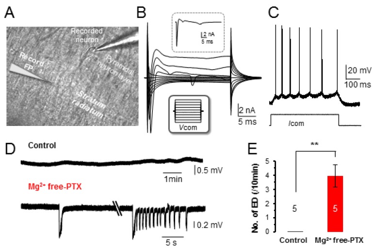
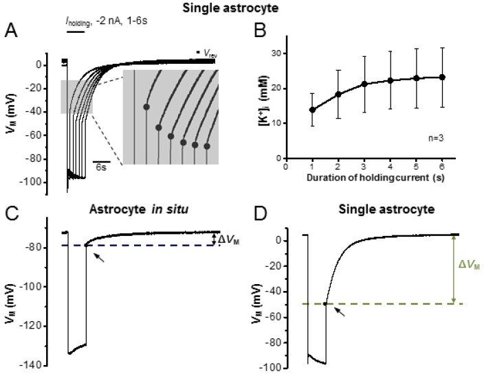
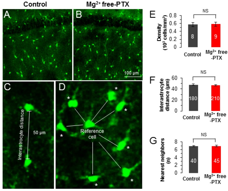
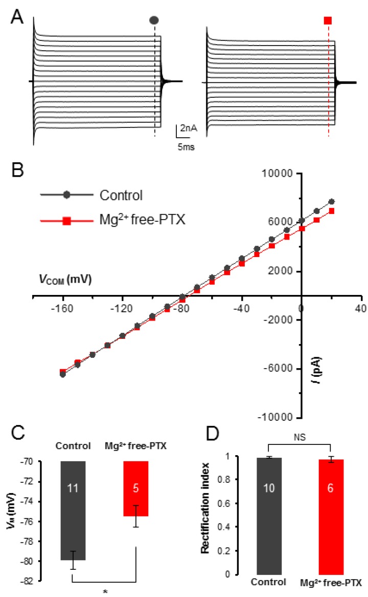
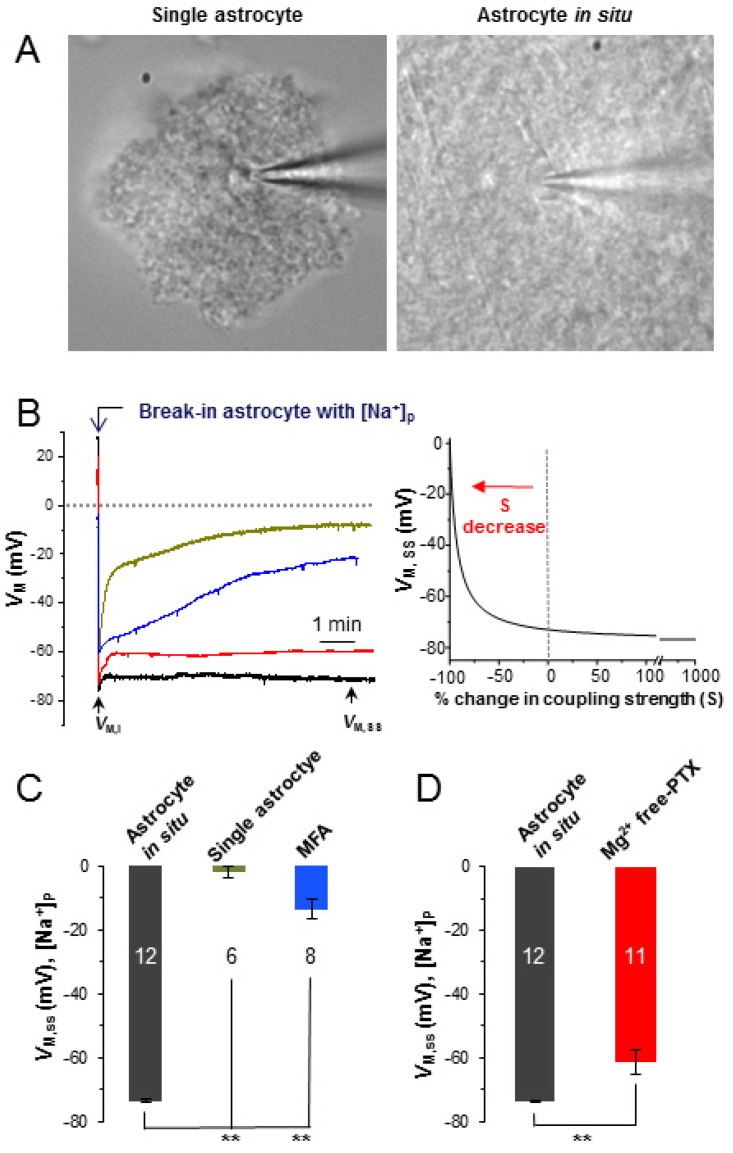
Astrocyte syncytial isopotentiality is a physiological mechanism resulting from a strong electrical coupling among astrocytes. We have previously shown that syncytial isopotentiality exists as a system-wide feature that coordinates astrocytes into a system for high efficient regulation of brain homeostasis. Neuronal activity is known to regulate gap junction coupling through alteration of extracellular ions and neurotransmitters. However, the extent to which epileptic neuronal activity impairs the syncytial isopotentiality is unknown. Here, the neuronal epileptiform bursts were induced in acute hippocampal slices by removal of Mg (Mg free) from bath solution and inhibition of γ-aminobutyric acid A (GABA) receptors by 100 µM picrotoxin (PTX). The change in syncytial coupling was monitored by using a K free-Na-containing electrode solution ([Na]) in the electrophysiological recording where the substitution of intracellular K by Na ions dissipates the physiological membrane potential (V) to ~0 mV in the recorded astrocyte. However, in a syncytial coupled astrocyte, the [Na] induced V loss can be compensated by the coupled astrocytes to a quasi-physiological membrane potential of ~73 mV. After short-term exposure to this experimental epileptic condition, a significant closure of syncytial coupling was indicated by a shift of the quasi-physiological membrane potential to -60 mV, corresponding to a 90% reduction of syncytial coupling strength. Consequently, the closure of syncytial coupling significantly decreased the ability of the syncytium for spatial redistribution of K ions. Altogether, our results show that epileptiform neuronal discharges weaken the strength of syncytial coupling and that in turn impairs the capacity of a syncytium for spatial redistribution of K ions.
星形胶质细胞同步等电位性是一种由星形胶质细胞之间强大的电耦合产生的生理机制。我们之前已经表明,同步等电位性作为一种全系统特征存在,它将星形胶质细胞协调成一个系统,以高效调节脑内稳态。已知神经元活动通过改变细胞外离子和神经递质来调节缝隙连接耦合。然而,癫痫性神经元活动损害同步等电位性的程度尚不清楚。在这里,通过从浴液中去除镁(无镁)并用100μM苦味毒(PTX)抑制γ-氨基丁酸A(GABA)受体,在急性海马切片中诱导神经元癫痫样爆发。在电生理记录中,通过使用无钾-含钠电极溶液([Na])监测同步耦合的变化,其中用钠离子替代细胞内钾会使记录的星形胶质细胞中的生理膜电位(V)消散至约0 mV。然而,在同步耦合的星形胶质细胞中,[Na]诱导的V损失可以由耦合的星形胶质细胞补偿至约73 mV的准生理膜电位。在短期暴露于这种实验性癫痫状态后,准生理膜电位向-60 mV的转变表明同步耦合显著关闭,这对应于同步耦合强度降低90%。因此,同步耦合的关闭显著降低了合胞体对钾离子进行空间重新分布的能力。总之,我们的结果表明,癫痫样神经元放电会削弱同步耦合的强度,进而损害合胞体对钾离子进行空间重新分布的能力。