Cheng Yuxin, Wang Ting, Lv Xin, Li Rutian, Yuan Ling, Shen Jie, Li Yan, Yan Tingting, Liu Baorui, Wang Lifeng
The Comprehensive Cancer Centre of Nanjing Drum Tower Hospital, Clinical College of Nanjing Medical University, Nanjing, People's Republic of China.
Department of Pathology, The Affiliated Drum Tower Hospital, Nanjing University Medical School, Nanjing, People's Republic of China.
Cancer Manag Res. 2020 Mar 19;12:2069-2078. doi: 10.2147/CMAR.S245425. eCollection 2020.
The expression of programmed cell death ligand 1(PD-L1) is related to the efficacy of immune checkpoint inhibitors on patients with non-small cell lung cancer (NSCLC), but tumor tissue (TT) samples are difficult to obtain, and initial TT samples are difficult to reflect the spatial-temporal heterogeneity. Therefore, we explored the feasibility of separating circulating tumor cells (CTCs) and detecting PD-L1 expression on CTCs.
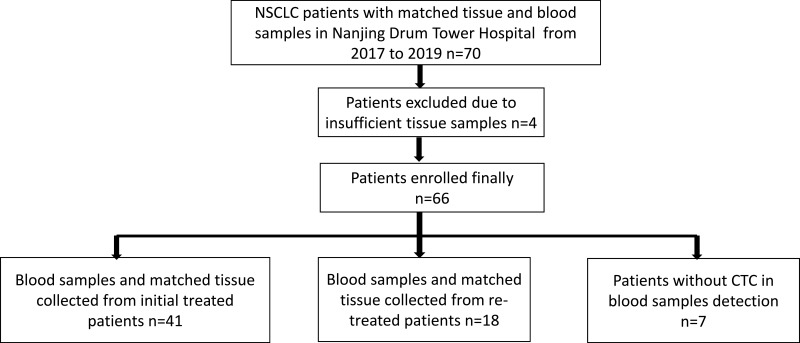
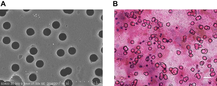
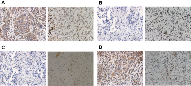
Peripheral blood specimens were sampled from 66 NSCLC patients, and CTCs were separated by membrane filtration based on size. For 59 patients with paired TT specimens, the expression of PD-L1 in their CTCs and TTs was determined using the immunohistochemistry and immunocytochemistry based on 28-8 antibody, respectively. The PD-L1 expression in TTs was set as a gold standard for calculation of sensitivity, specificity, consistency, positive predictive value (PPV), and negative predictive value (NPV), and the Cohen kappa coefficient for CTCs and paired TTs was calculated. In addition, the -test, Chi-square test, and Mann-Whitney -test were adopted to analyze the correlation of clinical pathological features and prognosis with PD-L1 expression.
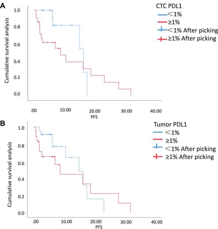
Sensitivity, specificity, concordance, PPV and NPV of detecting PD-L1 in CTCs of the 41 initial treated patients were 88.89%, 73.91%, 80%, 72.73% and 89.47%, respectively, and the Cohen kappa coefficient of CTC and paired TTs was 0.613. The univariate analysis of survival showed that the progression-free survival time of initial treated patients with positive PD-L1 expression was shorter than that of those with negative PD-L1 expression in CTCs or TTs (>0.05), and the positive PD-L1 expression in CTCs or TTs had nothing to do with age, sex, smoking status, histological type, and stage ( > 0.05).
The study confirms the feasibility of CTC PD-L1 detection in peripheral blood and lays a foundation for exploring real-time and individualized immunotherapy molecular biomarkers.
程序性细胞死亡配体1(PD-L1)的表达与免疫检查点抑制剂对非小细胞肺癌(NSCLC)患者的疗效相关,但肿瘤组织(TT)样本难以获取,且初始TT样本难以反映时空异质性。因此,我们探讨了分离循环肿瘤细胞(CTC)并检测CTC上PD-L1表达的可行性。
采集66例NSCLC患者的外周血标本,基于大小通过膜过滤分离CTC。对于59例有配对TT标本的患者,分别使用基于28-8抗体的免疫组织化学和免疫细胞化学方法测定其CTC和TT中PD-L1的表达。将TT中PD-L1的表达设定为计算敏感性、特异性、一致性、阳性预测值(PPV)和阴性预测值(NPV)的金标准,并计算CTC与配对TT的Cohen kappa系数。此外,采用t检验、卡方检验和Mann-Whitney U检验分析临床病理特征及预后与PD-L1表达的相关性。
41例初始治疗患者的CTC中检测PD-L1的敏感性、特异性、一致性、PPV和NPV分别为88.89%、73.91%、80%、72.73%和89.47%,CTC与配对TT的Cohen kappa系数为0.613。生存单因素分析显示,初始治疗患者中CTC或TT中PD-L1表达阳性者的无进展生存时间短于PD-L1表达阴性者(P>0.05),且CTC或TT中PD-L1表达阳性与年龄、性别、吸烟状态、组织学类型及分期无关(P>0.05)。
本研究证实了外周血中检测CTC的PD-L1的可行性,为探索实时、个体化免疫治疗分子生物标志物奠定了基础。