Cai Xingbo, Xu Yongqing, Yu Kaifu, He Xiaoqing, Luo Haotian, Duan Jiazhang, Wu Yipeng
From the The 920th Hospital of the Joint Logistic Support Force of the PLA, Kunming, Yunnan, China.
Ann Plast Surg. 2020 May;84(5S Suppl 3):S230-S234. doi: 10.1097/SAP.0000000000002362.
The aim of the study was to explore the feasibility and early effect of digital design combined with 3-dimensional (3D) printing technique in the transplantation of vascular pedicled iliac bone flap in the treatment of avascular necrosis of the femoral head.
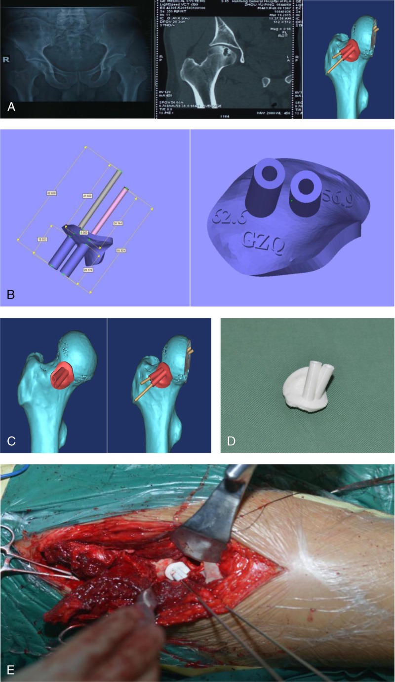
The navigation template was designed according to computed tomography scan and printed in 3D printing technique before operation, which was used to guide the localization and clearance of osteonecrosis of the femoral head in vascular pedicled iliac bone flap transplantation. In blank control group, 28 cases (32 hips) of osteonecrosis of the femoral head were treated with vascular pedicled iliac bone flap without the assistance of 3D navigation template from February 2002 to February 2009, including 19 males (21 hips) and 9 females (11 hips), with an average age of 37 years (range, 20-61 years). There were 12 cases of left hip, 16 cases of right hip, and 4 cases of double hip. According to the International Association of Bone Circulation staging, there were 8 hips in stage II B, 9 hips in stage II C, 8 hips in stage III B, and 7 hips in stage III C. In the experimental group, from February 2014 to June 2014, 15 patients (24 hips) with avascular necrosis of the femoral head were treated with vascular pedicled iliac bone flap with the aid of 3D navigation template. There were 11 males (17 hips) and 4 females (7 hips) with an average age of 38 years (range, 18-56 years). There were 2 cases of left hip, 4 cases of right hip, and 9 cases of double hip. According to the International Association of Bone Circulation staging, there were 5 hips in stage II B, 8 hips in stage II C, 6 hips in stage III B, and 5 hips in stage III C. The operation time, bleeding volume, and postoperative Harris score of the experimental group and the control group were statistically analyzed.
The incisions in both groups healed in the first stage, and there were no operation-related complications such as deep venous thrombosis and infection of lower extremities. All patients were followed up for 12 to 16 months (with an average of 14 months). On the second day after operation, X-ray and computed tomography showed that the necrotic focus of the femoral head and the surrounding sclerotic bone was completely removed, and the position of the vascular pedicled iliac bone flap was satisfactory and did not penetrate the articular surface. The iliac bone flap and bone graft achieved bony fusion. In the navigation template group, the mean ± SD operation time was 135.38 ± 9.49 minutes, the mean ± SD blood loss was 225.13 ± 13.41 mL, the mean ± SD postoperative Harris score was 89.53 ± 5.83, 12 hips were excellent, 10 hips were good, and 2 hips were moderate, whereas in the group without navigation template, the mean ± SD operation time was 151.00 ± 15.28 minutes, the mean ± SD blood loss was 283.56 ± 30.60 mL, the mean ± SD postoperative Harris score was 83.32 ± 3.75, 15 hips were excellent, 14 hips were good, and 3 hips were fair. By independent sample t test, there were significant differences in average operation time, average blood loss, and postoperative Harris score between the 2 groups (P < 0.05).
Compared with not using navigation template, vascular pedicled iliac bone flap combined with navigation template in the treatment of osteonecrosis of femoral head could locate the area of osteonecrosis of femoral head more accurately, shorten the time of operation, and reduce the amount of bleeding during operation. Postoperative hip joint function recovery was better, and the early effect was satisfactory.
本研究旨在探讨数字设计结合三维(3D)打印技术在带血管蒂髂骨瓣移植治疗股骨头缺血性坏死中的可行性及早期效果。
术前根据计算机断层扫描设计导航模板并采用3D打印技术打印,用于指导带血管蒂髂骨瓣移植治疗股骨头坏死时股骨头坏死灶的定位及清除。空白对照组为2002年2月至2009年2月28例(32髋)股骨头坏死患者,采用带血管蒂髂骨瓣治疗,未使用3D导航模板辅助,其中男性19例(21髋),女性9例(11髋),平均年龄37岁(范围20 - 61岁)。左髋12例,右髋16例,双侧髋4例。根据国际骨循环协会分期,II B期8髋,II C期9髋,III B期8髋,III C期7髋。实验组为2014年2月至2014年6月15例(24髋)股骨头缺血性坏死患者,采用带血管蒂髂骨瓣并借助3D导航模板治疗。男性11例(17髋),女性4例(7髋),平均年龄38岁(范围18 - 56岁)。左髋2例,右髋4例,双侧髋9例。根据国际骨循环协会分期,II B期5髋,II C期8髋,III B期6髋,III C期5髋。对实验组和对照组的手术时间、出血量及术后Harris评分进行统计学分析。
两组切口均一期愈合,未出现下肢深静脉血栓、感染等手术相关并发症。所有患者均随访12至16个月(平均14个月)。术后第二天,X线及计算机断层扫描显示股骨头坏死灶及周围硬化骨被完全清除,带血管蒂髂骨瓣位置满意,未穿透关节面。髂骨瓣与植骨实现骨性融合。导航模板组平均±标准差手术时间为135.38 ± 9.49分钟,平均±标准差失血量为225.13 ± 13.41 mL,平均±标准差术后Harris评分为89.53 ± 5.83,优12髋,良10髋,可2髋;而未使用导航模板组平均±标准差手术时间为151.00 ± 15.28分钟,平均±标准差失血量为283.56 ± 30.60 mL,平均±标准差术后Harris评分为83.32 ± 3.75,优15髋,良14髋,中3髋。经独立样本t检验,两组平均手术时间、平均出血量及术后Harris评分差异有统计学意义(P < 0.05)。
与不使用导航模板相比,带血管蒂髂骨瓣联合导航模板治疗股骨头坏死能更准确地定位股骨头坏死区域,缩短手术时间,减少术中出血量。术后髋关节功能恢复更好,早期效果满意。