Losurdo Michela, Davidsson Johan, Sköld Mattias K
Department of Neuroscience, Karolinska Institute, 171 77 Stockholm, Sweden.
Department of Molecular Medicine, Università degli Studi di Pavia, 27100 Pavia, Italy.
Brain Sci. 2020 Apr 10;10(4):229. doi: 10.3390/brainsci10040229.
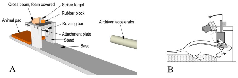
Traumatic brain injury (TBI) commonly results in primary diffuse axonal injury (DAI) and associated secondary injuries that evolve through a cascade of pathological mechanisms. We aim at assessing how myelin and oligodendrocytes react to head angular-acceleration-induced TBI in a previously described model. This model induces axonal injuries visible by amyloid precursor protein (APP) expression, predominantly in the corpus callosum and its borders. Brain tissue from a total of 27 adult rats was collected at 24 h, 72 h and 7 d post-injury. Coronal sections were prepared for immunohistochemistry and RNAscope to investigate DAI and myelin changes (APP, MBP, Rip), oligodendrocyte lineage cell loss (Olig2), oligodendrocyte progenitor cells (OPCs) (NG2, PDGFRa) and neuronal stress (HSP70, ATF3). Oligodendrocytes and OPCs numbers (expressed as percentage of positive cells out of total number of cells) were measured in areas with high APP expression. Results showed non-statistically significant trends with a decrease in oligodendrocyte lineage cells and an increase in OPCs. Levels of myelination were mostly unaltered, although Rip expression differed significantly between sham and injured animals in the frontal brain. Neuronal stress markers were induced at the dorsal cortex and habenular nuclei. We conclude that rotational injury induces DAI and neuronal stress in specific areas. We noticed indications of oligodendrocyte death and regeneration without statistically significant changes at the timepoints measured, despite indications of axonal injuries and neuronal stress. This might suggest that oligodendrocytes are robust enough to withstand this kind of trauma, knowledge important for the understanding of thresholds for cell injury and post-traumatic recovery potential.
创伤性脑损伤(TBI)通常会导致原发性弥漫性轴索损伤(DAI)以及相关的继发性损伤,这些损伤会通过一系列病理机制不断发展。我们旨在评估在先前描述的模型中,髓鞘和少突胶质细胞对头部角加速度诱导的TBI如何做出反应。该模型可诱导出以淀粉样前体蛋白(APP)表达可见的轴索损伤,主要位于胼胝体及其边界。在损伤后24小时、72小时和7天收集了总共27只成年大鼠的脑组织。制备冠状切片用于免疫组织化学和RNAscope检测,以研究DAI和髓鞘变化(APP、MBP、Rip)、少突胶质细胞谱系细胞丢失(Olig2)、少突胶质前体细胞(OPCs)(NG2、PDGFRa)和神经元应激(HSP70、ATF3)。在APP高表达区域测量少突胶质细胞和OPCs的数量(以阳性细胞占细胞总数的百分比表示)。结果显示少突胶质细胞谱系细胞减少和OPCs增加的趋势无统计学意义。髓鞘形成水平大多未改变,尽管在额叶中,假手术组和损伤组动物之间Rip表达存在显著差异。在背侧皮质和缰核诱导出神经元应激标志物。我们得出结论,旋转损伤会在特定区域诱导DAI和神经元应激。我们注意到少突胶质细胞死亡和再生的迹象,尽管存在轴索损伤和神经元应激的迹象,但在所测量的时间点上无统计学显著变化。这可能表明少突胶质细胞足够强健,能够承受这种创伤,这一知识对于理解细胞损伤阈值和创伤后恢复潜力很重要。