Sanders-Brown Center on Aging, University of Kentucky, Room 433, Sanders-Brown Bldg., 800 S. Limestone Street, Lexington, KY, 40536, USA.
Department of Neuroscience, University of Kentucky, Lexington, KY, 40536, USA.
J Neuroinflammation. 2020 Apr 14;17(1):115. doi: 10.1186/s12974-020-01800-w.
Older-age individuals are at the highest risk for disability from a traumatic brain injury (TBI). Astrocytes are the most numerous glia in the brain, necessary for brain function, yet there is little known about unique responses of astrocytes in the aged-brain following TBI.
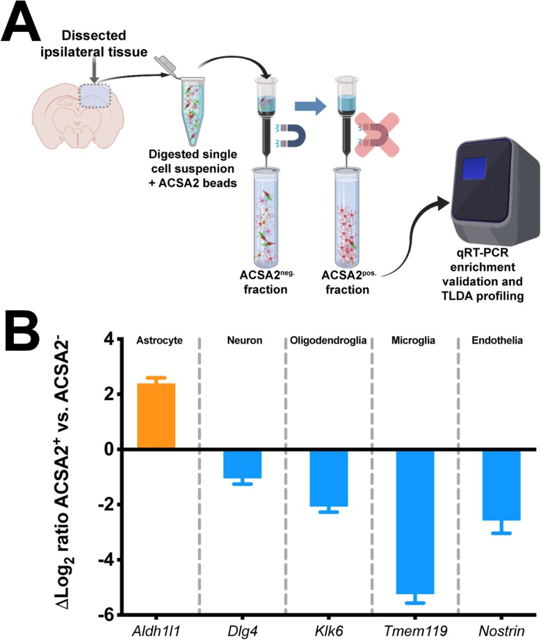
Our approach examined astrocytes in young adult, 4-month-old, versus aged, 18-month-old mice, at 1, 3, and 7 days post-TBI. We selected these time points to span the critical period in the transition from acute injury to presumably irreversible tissue damage and disability. Two approaches were used to define the astrocyte contribution to TBI by age interaction: (1) tissue histology and morphological phenotyping, and (2) transcriptomics on enriched astrocytes from the injured brain.
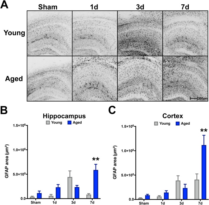
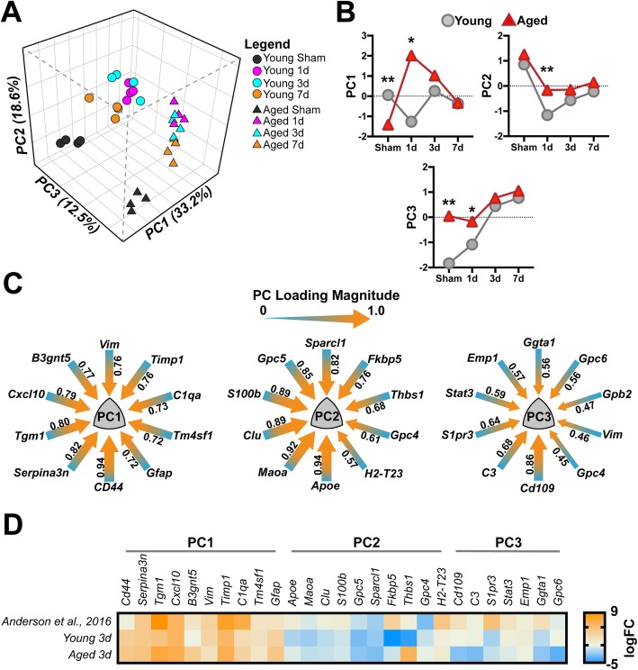
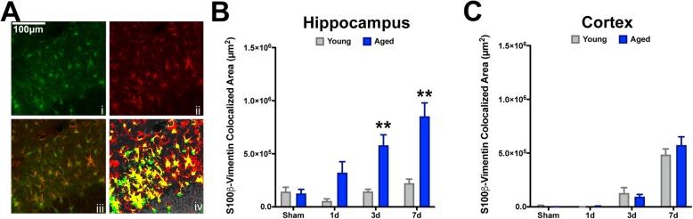
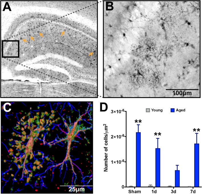
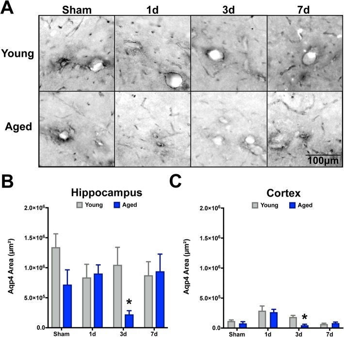
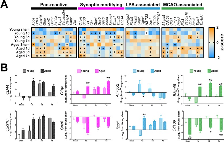
Aging was found to have a profound effect on the TBI-induced loss of astrocyte function needed for maintaining water transport and edema-namely, aquaporin-4. The aged brain also demonstrated a progressive exacerbation of astrogliosis as a function of time after injury. Moreover, clasmatodendrosis, an underrecognized astrogliopathy, was found to be significantly increased in the aged brain, but not in the young brain. As a function of TBI, we observed a transitory refraction in the number of these astrocytes, which rebounded by 7 days post-injury in the aged brain. Transcriptomic data demonstrated disproportionate changes in genes attributed to reactive astrocytes, inflammatory response, complement pathway, and synaptic support in aged mice following TBI compared to young mice. Additionally, our data highlight that TBI did not evoke a clear alignment with the previously defined "A1/A2" dichotomy of reactive astrogliosis.
Overall, our findings point toward a progressive phenotype of aged astrocytes following TBI that we hypothesize to be maladaptive, shedding new insights into potentially modifiable astrocyte-specific mechanisms that may underlie increased fragility of the aged brain to trauma.
年龄较大的个体因创伤性脑损伤(TBI)而残疾的风险最高。星形胶质细胞是大脑中数量最多的神经胶质细胞,对大脑功能至关重要,但人们对 TBI 后老年大脑中星形胶质细胞的独特反应知之甚少。
我们的方法研究了年轻成年(4 个月大)和老年(18 个月大)小鼠在 TBI 后 1、3 和 7 天的星形胶质细胞。我们选择这些时间点来跨越从急性损伤到可能不可逆转的组织损伤和残疾的关键过渡时期。使用两种方法来定义年龄对 TBI 相互作用的星形胶质细胞贡献:(1)组织组织学和形态表型,以及(2)从受伤大脑中富集的星形胶质细胞的转录组学。
研究发现,衰老对维持水转运和水肿所需的星形胶质细胞功能的 TBI 诱导丧失有深远的影响,即水通道蛋白-4。老年大脑还表现出随着损伤后时间的推移,星形胶质细胞增生逐渐加剧。此外,发现少突胶质细胞病(一种未被充分认识的星形胶质细胞病)在老年大脑中显著增加,但在年轻大脑中没有增加。作为 TBI 的一个功能,我们观察到这些星形胶质细胞数量的短暂折射,在老年大脑中,这种折射在损伤后 7 天反弹。转录组学数据表明,与年轻小鼠相比,TBI 后老年小鼠与反应性星形胶质细胞、炎症反应、补体途径和突触支持相关的基因发生了不成比例的变化。此外,我们的数据还表明,TBI 并没有明确指向以前定义的反应性星形胶质细胞的“A1/A2”二分法。
总的来说,我们的研究结果表明,TBI 后老年星形胶质细胞的表型逐渐发生变化,我们假设这是一种适应不良的变化,为潜在的可改变的星形胶质细胞特异性机制提供了新的见解,这些机制可能是导致老年大脑对创伤更加脆弱的原因。