• 文献检索
  • 文档翻译
  • 深度研究
  • 学术资讯
  • Suppr Zotero 插件Zotero 插件
  • 邀请有礼
  • 套餐&价格
  • 历史记录
应用&插件
Suppr Zotero 插件Zotero 插件浏览器插件Mac 客户端Windows 客户端微信小程序
定价
高级版会员购买积分包购买API积分包
服务
文献检索文档翻译深度研究API 文档MCP 服务
关于我们
关于 Suppr公司介绍联系我们用户协议隐私条款
关注我们

Suppr 超能文献

核心技术专利:CN118964589B侵权必究
粤ICP备2023148730 号-1Suppr @ 2026

文献检索

告别复杂PubMed语法,用中文像聊天一样搜索,搜遍4000万医学文献。AI智能推荐,让科研检索更轻松。

立即免费搜索

文件翻译

保留排版,准确专业,支持PDF/Word/PPT等文件格式,支持 12+语言互译。

免费翻译文档

深度研究

AI帮你快速写综述,25分钟生成高质量综述,智能提取关键信息,辅助科研写作。

立即免费体验

基于定量 MRI 的骨髓炎水肿和脂肪的组织学分析。

Histographic analysis of oedema and fat in inflamed bone marrow based on quantitative MRI.

机构信息

Centre for Medical Imaging, University College London, London, UK.

Arthritis Research UK Centre for Adolescent Rheumatology, University College London, London, UK.

出版信息

Eur Radiol. 2020 Sep;30(9):5099-5109. doi: 10.1007/s00330-020-06785-x. Epub 2020 Apr 14.

DOI:10.1007/s00330-020-06785-x
PMID:32291499
原文链接:https://pmc.ncbi.nlm.nih.gov/articles/PMC7431434/
Abstract

OBJECTIVE

To demonstrate proof-of-concept for a quantitative MRI method using histographic analysis to assess bone marrow oedema and fat metaplasia in the sacroiliac joints.

MATERIALS AND METHODS

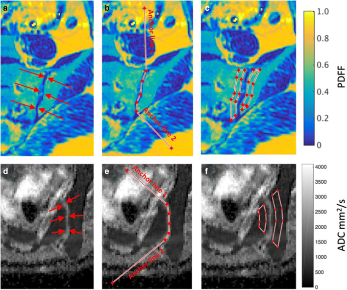
Fifty-three adolescents aged 12-23 with known or suspected sacroiliitis were prospectively recruited and underwent quantitative MRI (qMRI) scans, consisting of chemical shift-encoded (at 3 T) and diffusion-weighted imaging (at 1.5 T), plus conventional MRI (at 1.5 T) and clinical assessment. qMRI scans produced proton-density fat fraction (PDFF) and apparent diffusion coefficient (ADC) maps of the sacroiliac joints (SIJs), which were analysed using an in-house software tool enabling partially automated ROI definition and histographic analysis. Logistic regression and receiver operating characteristic (ROC) analyses assessed the predictive performance of ADC- and PDFF-based parameters in identifying active inflammation (oedema) and structural damage (fat metaplasia).

RESULTS

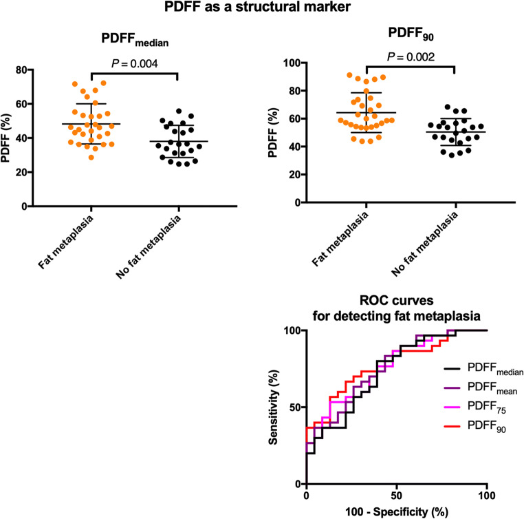
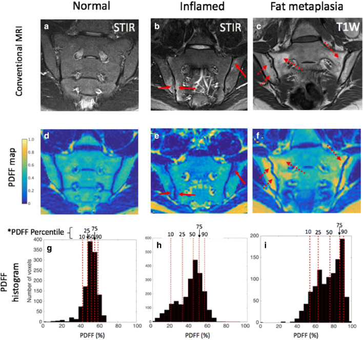
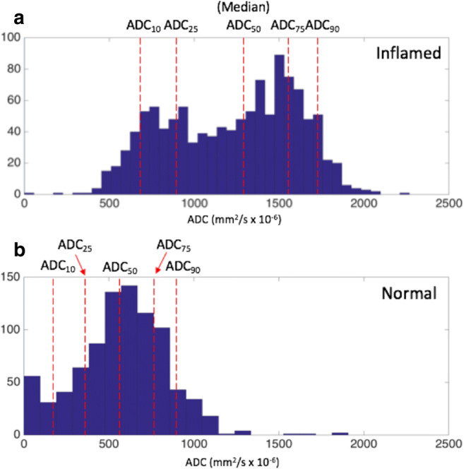
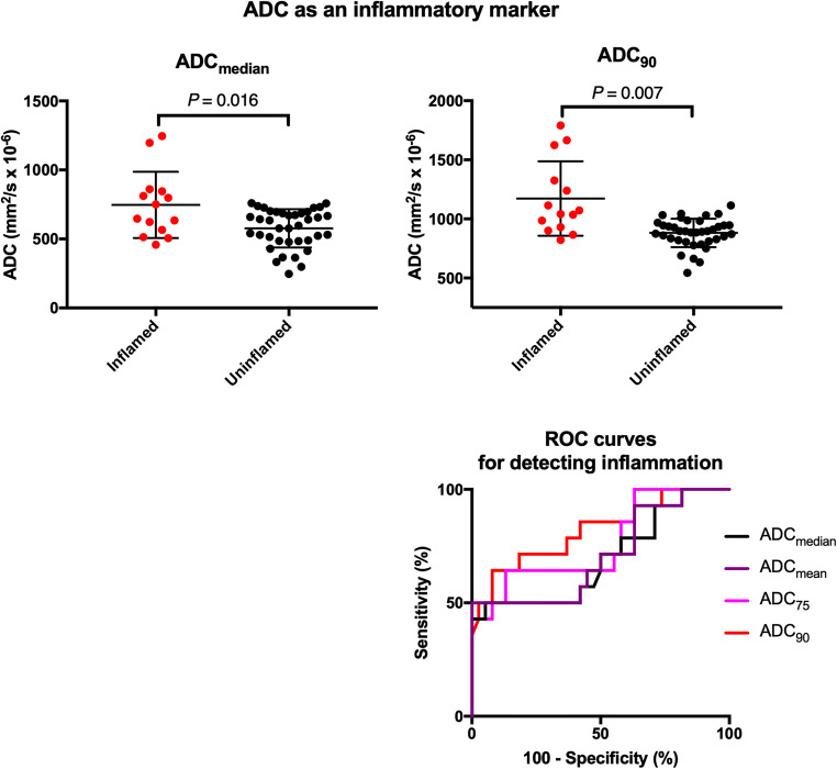
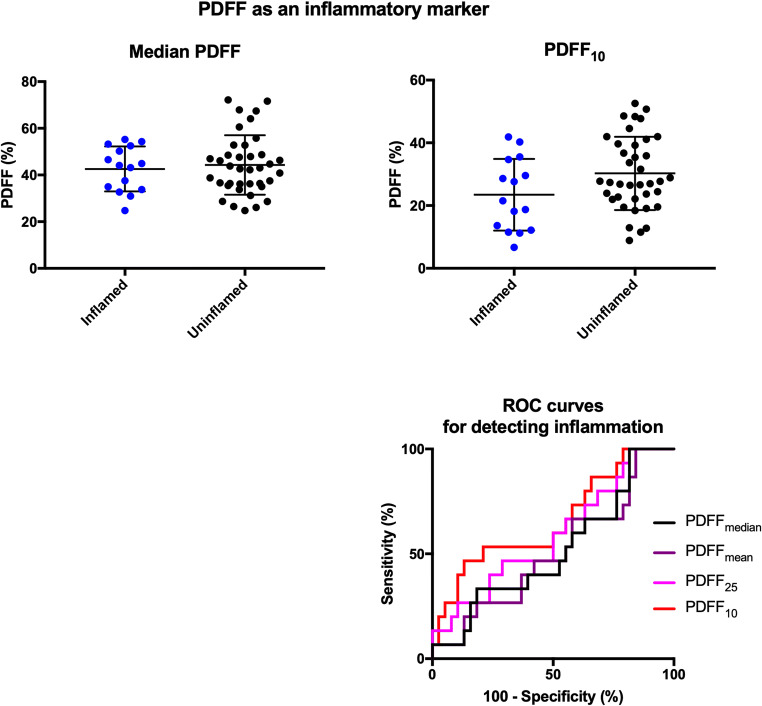
ADC-based parameters were associated with increased odds of oedema (all p < 0.05); ROC-AUC was higher for histographic parameters representing the upper end of the ADC distribution than for simple averages. Similarly, PDFF-based parameters were associated with increased odds of fat metaplasia (all p < 0.05); ROC area-under-the-curve was higher for histographic parameters representing the upper end of the PDFF distribution than for simple averages. Both ADC- and PDFF-based histographic parameters demonstrated excellent inter- and intra-observer agreement (ICC > 0.9).

CONCLUSIONS

ADC-based parameters can differentiate patients with bone marrow oedema from those without, whilst PDFF-based parameters can differentiate patients with fat metaplasia from those without. Histographic analysis might improve performance compared with simple averages such as the mean and median and offers excellent agreement within and between observers.

KEY POINTS

• Quantitative MRI with histographic analysis can identify bone marrow oedema (an active inflammatory lesion) and fat metaplasia (a 'chronic' inflammatory lesion) in patients with spondyloarthritis. • The use of histographic analysis might improve the performance of quantitative MRI for detecting bone marrow oedema and fat metaplasia compared with simple averages such as the mean and median. • Bone marrow oedema and fat metaplasia are known to be of diagnostic and prognostic significance, and the proposed method could support clinical decisions around biologic (and other) therapies in spondyloarthritis.

摘要

目的

通过使用直方图分析来证明一种定量 MRI 方法的概念验证,以评估骶髂关节中的骨髓水肿和脂肪化生。

材料与方法

53 名年龄在 12-23 岁之间的已知或疑似骶髂关节炎的青少年前瞻性入组并接受定量 MRI(qMRI)扫描,包括化学位移编码(3T)和弥散加权成像(1.5T),加上常规 MRI(1.5T)和临床评估。qMRI 扫描产生骶髂关节(SIJ)的质子密度脂肪分数(PDFF)和表观扩散系数(ADC)图,使用内部软件工具对其进行分析,该工具可实现部分自动 ROI 定义和直方图分析。逻辑回归和受试者工作特征(ROC)分析评估了基于 ADC 和 PDFF 的参数在识别活跃炎症(水肿)和结构损伤(脂肪化生)方面的预测性能。

结果

基于 ADC 的参数与水肿的可能性增加相关(均 p<0.05);代表 ADC 分布上限的直方图参数的 ROC-AUC 高于简单平均值。同样,基于 PDFF 的参数与脂肪化生的可能性增加相关(均 p<0.05);代表 PDFF 分布上限的直方图参数的 ROC 曲线下面积高于简单平均值。基于 ADC 和 PDFF 的直方图参数均表现出极好的观察者内和观察者间一致性(ICC>0.9)。

结论

基于 ADC 的参数可区分骨髓水肿患者与无骨髓水肿患者,而基于 PDFF 的参数可区分脂肪化生患者与无脂肪化生患者。与平均值(如均值和中位数)相比,直方图分析可能会提高性能,并在观察者内和观察者间提供极好的一致性。

关键点

• 定量 MRI 结合直方图分析可识别出患有脊柱关节炎的患者中的骨髓水肿(活跃炎症病变)和脂肪化生(慢性炎症病变)。• 与平均值(如均值和中位数)相比,直方图分析可能会提高定量 MRI 检测骨髓水肿和脂肪化生的性能。• 骨髓水肿和脂肪化生具有诊断和预后意义,所提出的方法可以支持脊柱关节炎中生物(和其他)治疗的临床决策。

https://cdn.ncbi.nlm.nih.gov/pmc/blobs/03bc/7431434/a3ca672a2a04/330_2020_6785_Fig6_HTML.jpg
https://cdn.ncbi.nlm.nih.gov/pmc/blobs/03bc/7431434/74791e6223ce/330_2020_6785_Fig1_HTML.jpg
https://cdn.ncbi.nlm.nih.gov/pmc/blobs/03bc/7431434/85c878b33c44/330_2020_6785_Fig2_HTML.jpg
https://cdn.ncbi.nlm.nih.gov/pmc/blobs/03bc/7431434/45b81c02900d/330_2020_6785_Fig3_HTML.jpg
https://cdn.ncbi.nlm.nih.gov/pmc/blobs/03bc/7431434/ad52aed39d9d/330_2020_6785_Fig4_HTML.jpg
https://cdn.ncbi.nlm.nih.gov/pmc/blobs/03bc/7431434/0fad10a133c4/330_2020_6785_Fig5_HTML.jpg
https://cdn.ncbi.nlm.nih.gov/pmc/blobs/03bc/7431434/a3ca672a2a04/330_2020_6785_Fig6_HTML.jpg

相似文献

1
Histographic analysis of oedema and fat in inflamed bone marrow based on quantitative MRI.基于定量 MRI 的骨髓炎水肿和脂肪的组织学分析。
Eur Radiol. 2020 Sep;30(9):5099-5109. doi: 10.1007/s00330-020-06785-x. Epub 2020 Apr 14.
2
Quantification of bone marrow oedema and fat metaplasia in sacroiliac joints in spondyloarthritis patients using histographic magnetic resonance imaging analysis.采用组织直方图磁共振成像分析定量评估脊柱关节炎患者骶髂关节骨髓水肿和脂肪化生。
Clin Exp Rheumatol. 2022 Jul;40(7):1385-1392. doi: 10.55563/clinexprheumatol/e3b8c9. Epub 2021 Sep 28.
3
Proton density fat fraction (PDFF) maps aid fat metaplasia evaluation in the sacroiliac joints in additional to T1WI: Improved diagnostic accuracy in axial spondyloarthritis.质子密度脂肪分数(PDFF)图谱有助于在骶髂关节中除 T1WI 以外评估脂肪化生:在轴向脊椎关节炎中提高诊断准确性。
Eur J Radiol. 2022 Dec;157:110569. doi: 10.1016/j.ejrad.2022.110569. Epub 2022 Oct 22.
4
Quantification of Fat Metaplasia in the Sacroiliac Joints of Patients With Axial Spondyloarthritis by Chemical Shift-Encoded MRI: A Diagnostic Trial.化学位移编码 MRI 定量评估强直性脊柱炎患者骶髂关节脂肪化生:一项诊断性试验。
Front Immunol. 2022 Jan 18;12:811672. doi: 10.3389/fimmu.2021.811672. eCollection 2021.
5
Simultaneous Quantification of Bone Edema/Adiposity and Structure in Inflamed Bone Using Chemical Shift-Encoded MRI in Spondyloarthritis.应用化学位移编码 MRI 技术对脊柱关节炎患者炎症骨的水肿/脂肪含量和结构进行同时定量分析。
Magn Reson Med. 2018 Feb;79(2):1031-1042. doi: 10.1002/mrm.26729. Epub 2017 Jun 6.
6
Scoring magnetic resonance imaging (MRI) inflammation and structural lesions in sacroiliac joints of patients with axial spondyloarthritis: assessment of all MRI slices of the cartilaginous compartment versus standardized six or five slices.评估中轴型脊柱关节炎患者骶髂关节的磁共振成像(MRI)炎症和结构病变:评估软骨间隙的所有 MRI 层面与标准的 6 或 5 个层面。
Scand J Rheumatol. 2020 May;49(3):200-209. doi: 10.1080/03009742.2019.1675184. Epub 2019 Dec 17.
7
Synthetic MRI in the detection and quantitative evaluation of sacroiliac joint lesions in axial spondyloarthritis.磁共振合成技术在轴性脊柱关节炎中骶髂关节病变的检测和定量评估中的应用。
Front Immunol. 2022 Sep 26;13:1000314. doi: 10.3389/fimmu.2022.1000314. eCollection 2022.
8
Axial involvement in patients with early peripheral spondyloarthritis: a prospective MRI study of sacroiliac joints and spine.早期外周型脊柱关节炎患者的轴向受累:骶髂关节和脊柱的前瞻性 MRI 研究。
Ann Rheum Dis. 2021 Jan;80(1):103-108. doi: 10.1136/annrheumdis-2020-218480. Epub 2020 Oct 28.
9
Comparison of proton density fat fraction, simultaneous R2*, and apparent diffusion coefficient for assessment of focal vertebral bone marrow lesions.质子密度脂肪分数、同时 R2*和表观扩散系数在评估局灶性椎体骨髓病变中的比较。
Clin Radiol. 2020 Feb;75(2):123-130. doi: 10.1016/j.crad.2019.09.141. Epub 2019 Oct 31.
10
MRI of the sacroiliac joints in athletes: recognition of non-specific bone marrow oedema by semi-axial added to standard semi-coronal scans.运动员的骶髂关节 MRI:通过半轴向加标准半冠状扫描识别非特异性骨髓水肿。
Rheumatology (Oxford). 2020 Jun 1;59(6):1381-1390. doi: 10.1093/rheumatology/kez458.

引用本文的文献

1
Application of apparent diffusion coefficient of extraocular muscles from diffusion tensor imaging scanning in the assessment of disease activity of thyroid eye disease.扩散张量成像扫描测量的眼外肌表观扩散系数在甲状腺眼病疾病活动度评估中的应用
BMC Endocr Disord. 2024 Dec 24;24(1):276. doi: 10.1186/s12902-024-01818-8.
2
Applications of Diffusion-Weighted MRI to the Musculoskeletal System.扩散加权 MRI 在肌肉骨骼系统中的应用。
J Magn Reson Imaging. 2024 Feb;59(2):376-396. doi: 10.1002/jmri.28870. Epub 2023 Jul 21.
3
Quantitative magnetic resonance imaging (qMRI) in axial spondyloarthritis.

本文引用的文献

1
Performance of magnetic resonance imaging in the diagnosis of axial spondyloarthritis: a systematic literature review.磁共振成像在轴性脊柱关节炎诊断中的性能:系统文献回顾。
Rheumatology (Oxford). 2019 Nov 1;58(11):1955-1965. doi: 10.1093/rheumatology/kez172.
2
Recommendations for acquisition and interpretation of MRI of the spine and sacroiliac joints in the diagnosis of axial spondyloarthritis in the UK.英国推荐的用于诊断中轴型脊柱关节炎的脊柱和骶髂关节 MRI 采集和解读的指南。
Rheumatology (Oxford). 2019 Oct 1;58(10):1831-1838. doi: 10.1093/rheumatology/kez173.
3
Simultaneous Quantification of Bone Edema/Adiposity and Structure in Inflamed Bone Using Chemical Shift-Encoded MRI in Spondyloarthritis.
轴性脊柱关节炎的定量磁共振成像(qMRI)。
Br J Radiol. 2023 Mar 1;96(1144):20220675. doi: 10.1259/bjr.20220675. Epub 2023 Jan 25.
4
Water Fraction Ratio of the Sacroiliac Joint Subchondral Bone Marrow in Patients with Ankylosing Spondylitis Predicts the Degree of Disease Activity.强直性脊柱炎患者骶髂关节软骨下骨髓的水分分数比可预测疾病活动程度。
Diagnostics (Basel). 2022 Nov 17;12(11):2842. doi: 10.3390/diagnostics12112842.
5
Baseline Severity of Sacroiliitis and Extensive Fat Metaplasia Predicts the Progression of Backfill at the Sacroiliac Joint in Patients With Axial Spondyloarthritis.基线骶髂关节炎严重程度和广泛脂肪化生可预测中轴型脊柱关节炎患者骶髂关节填充的进展。
Front Immunol. 2022 Jun 27;13:887470. doi: 10.3389/fimmu.2022.887470. eCollection 2022.
6
Proton density fat fraction: magnetic resonance imaging applications beyond the liver.质子密度脂肪分数:肝脏以外的磁共振成像应用。
Diagn Interv Radiol. 2022 Jan;28(1):83-91. doi: 10.5152/dir.2021.21845.
7
Quantification of Fat Metaplasia in the Sacroiliac Joints of Patients With Axial Spondyloarthritis by Chemical Shift-Encoded MRI: A Diagnostic Trial.化学位移编码 MRI 定量评估强直性脊柱炎患者骶髂关节脂肪化生:一项诊断性试验。
Front Immunol. 2022 Jan 18;12:811672. doi: 10.3389/fimmu.2021.811672. eCollection 2021.
8
Main Diagnostic Pitfalls in Reading the Sacroiliac Joints on MRI.MRI 骶髂关节读片的主要诊断陷阱
Diagnostics (Basel). 2021 Oct 28;11(11):2001. doi: 10.3390/diagnostics11112001.
9
Oedema on STIR modified the effect of amoxicillin as treatment for chronic low back pain with Modic changes-subgroup analysis of a randomized trial.短TI反转恢复序列(STIR)上的水肿改变了阿莫西林作为治疗伴有Modic改变的慢性下腰痛的疗效——一项随机试验的亚组分析
Eur Radiol. 2021 Jun;31(6):4285-4297. doi: 10.1007/s00330-020-07542-w. Epub 2020 Nov 27.
应用化学位移编码 MRI 技术对脊柱关节炎患者炎症骨的水肿/脂肪含量和结构进行同时定量分析。
Magn Reson Med. 2018 Feb;79(2):1031-1042. doi: 10.1002/mrm.26729. Epub 2017 Jun 6.
4
The Use of Magnetic Resonance Imaging in Axial Spondyloarthritis: Time to Bridge the Gap Between Radiologists and Rheumatologists.磁共振成像在轴性脊柱关节炎中的应用:弥合放射科医生与风湿病科医生之间差距的时候了。
J Rheumatol. 2017 Jun;44(6):780-785. doi: 10.3899/jrheum.161337. Epub 2017 Apr 1.
5
Axial spondyloarthritis.中轴型脊柱关节炎。
Lancet. 2017 Jul 1;390(10089):73-84. doi: 10.1016/S0140-6736(16)31591-4. Epub 2017 Jan 20.
6
Diffusion-weighted imaging is a sensitive biomarker of response to biologic therapy in enthesitis-related arthritis.扩散加权成像在附着点炎相关关节炎中是生物治疗反应的敏感生物标志物。
Rheumatology (Oxford). 2017 Mar 1;56(3):399-407. doi: 10.1093/rheumatology/kew429.
7
Can we use structural lesions seen on MRI of the sacroiliac joints reliably for the classification of patients according to the ASAS axial spondyloarthritis criteria? Data from the DESIR cohort.我们能否可靠地根据 ASAS 轴性脊柱关节炎标准,使用骶髂关节 MRI 上观察到的结构损伤对患者进行分类?来自 DESIR 队列的数据。
Ann Rheum Dis. 2017 Feb;76(2):392-398. doi: 10.1136/annrheumdis-2016-209405. Epub 2016 Aug 4.
8
Association of the apparent diffusion coefficient with maturity in adolescent sacroiliac joints.青少年骶髂关节表观扩散系数与成熟度的相关性
J Magn Reson Imaging. 2016 Sep;44(3):556-64. doi: 10.1002/jmri.25209. Epub 2016 Feb 21.
9
Defining active sacroiliitis on MRI for classification of axial spondyloarthritis: update by the ASAS MRI working group.MRI 中活动性骶髂关节炎的定义用于强直性脊柱炎的分类:ASAS MRI 工作组的更新。
Ann Rheum Dis. 2016 Nov;75(11):1958-1963. doi: 10.1136/annrheumdis-2015-208642. Epub 2016 Jan 14.
10
A diffusion-based quantification technique for assessment of sacroiliitis in adolescents with enthesitis-related arthritis.一种基于扩散的量化技术,用于评估患有附着点炎相关关节炎的青少年的骶髂关节炎。
Br J Radiol. 2016;89(1059):20150775. doi: 10.1259/bjr.20150775. Epub 2015 Dec 7.