Sofia Soccorsa, Boccatonda Andrea, Montanari Marco, Spampinato Michele, D'ardes Damiano, Cocco Giulio, Accogli Esterita, Cipollone Francesco, Schiavone Cosima
Emergency Department, Ospedale Maggiore AUSL Bologna, Bologna, Italy.
Internistic Ultrasound Unit, SS Annunziata Hospital, "G. d'Annunzio" University, via dei Vestini, 66100, Chieti, Italy.
J Ultrasound. 2020 Jun;23(2):217-221. doi: 10.1007/s40477-020-00458-7. Epub 2020 Apr 16.
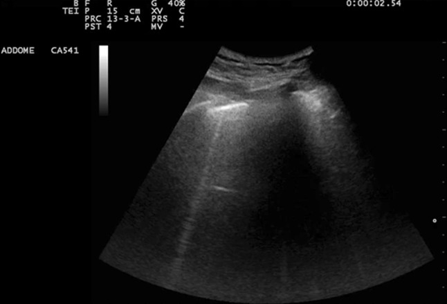
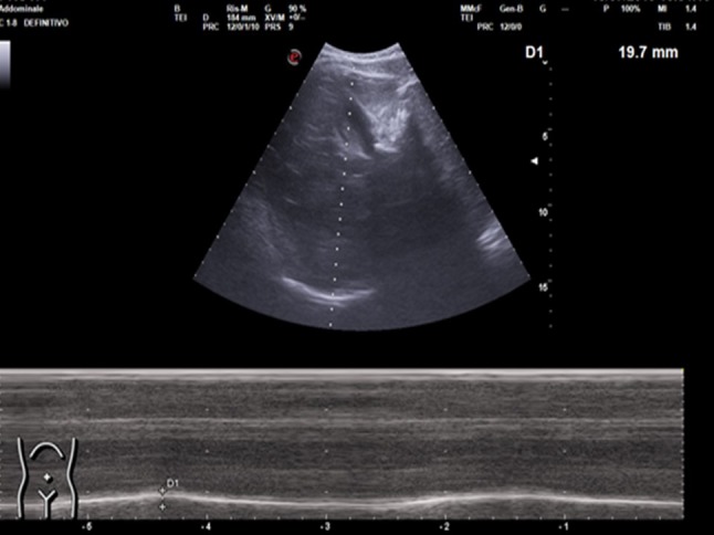
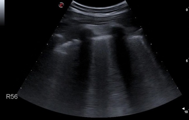
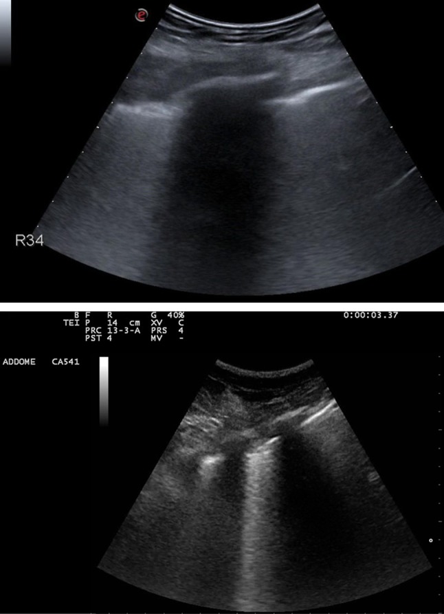
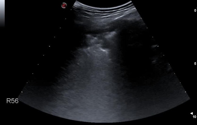
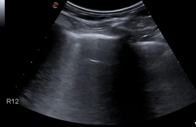
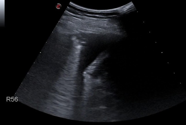
Thoracic ultrasound seems to adapt to the screening for lung involvement of patients with suspected or ascertained SARS-COVID-19 infection due to its characteristics of easy applicability. It can be also a relevant method in monitoring patients. B lines are early finding of COVID-19, even in mild-symptomatic subjects; in the most serious cases such as pre-ARDS or ARDS, the B lines end up filling the ultrasound image almost completely, until it merges, so as to create a single hyperechoic image named as "white lung", with distortion and irregularity of the pleural line. In advanced stage, lung consolidations are present, representing pulmonary pathological areas that are no longer normally ventilated.
由于易于应用的特点,胸部超声似乎适用于对疑似或确诊的SARS-CoV-2感染患者的肺部受累情况进行筛查。它也可以是监测患者的一种相关方法。B线是新冠病毒病的早期表现,即使在症状轻微的患者中也是如此;在最严重的病例中,如急性呼吸窘迫综合征前期或急性呼吸窘迫综合征,B线最终几乎完全充满超声图像,直至融合,从而形成一个名为“白肺”的单一高回声图像,同时伴有胸膜线的扭曲和不规则。在疾病晚期,会出现肺实变,代表不再正常通气的肺部病理区域。