Guangdong Provincial Key Laboratory of Malignant Tumor Epigenetics and Gene Regulation, Sun Yat-sen Memorial Hospital, Sun Yat-sen University, Guangzhou, Guangdong Province, China.
Department of Pancreatobiliary Surgery, Sun Yat-sen Memorial Hospital, Sun Yat-sen University, Guangzhou, Guangdong Province, China.
Theranostics. 2020 Apr 6;10(11):5029-5047. doi: 10.7150/thno.42440. eCollection 2020.
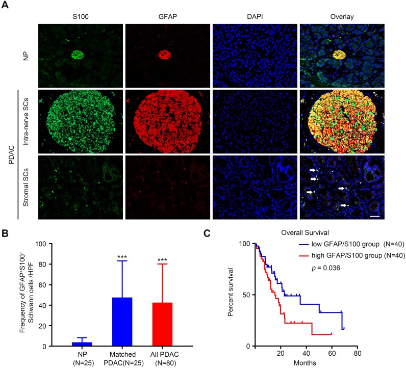
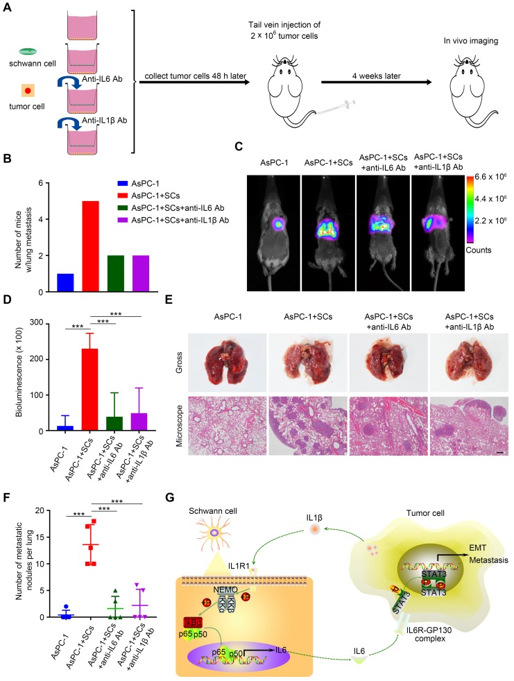
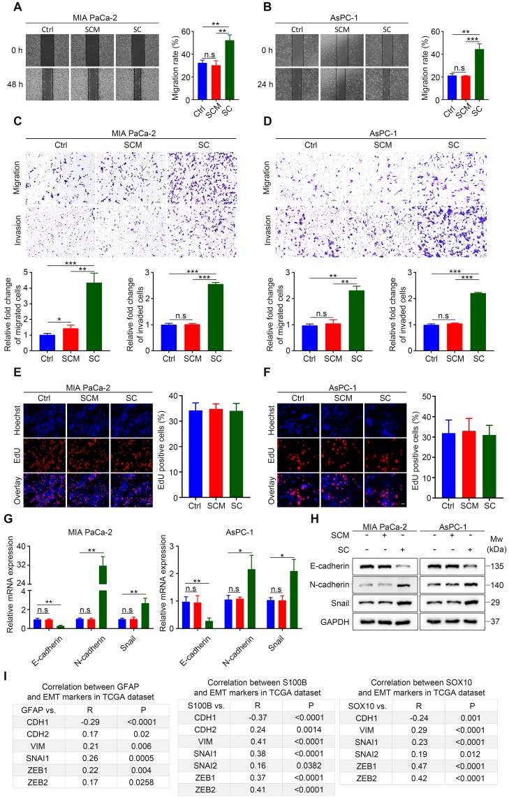
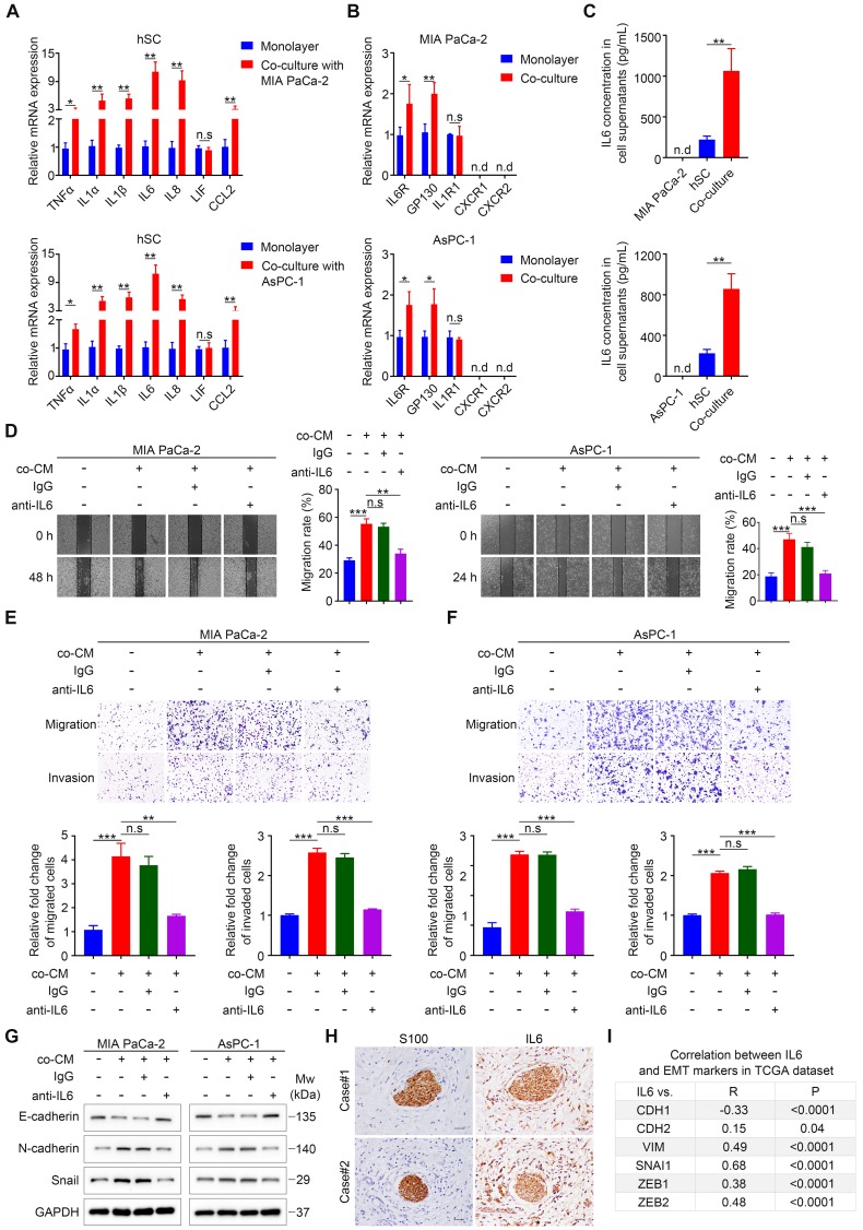
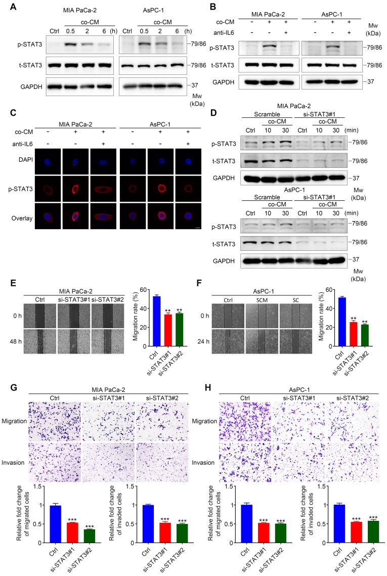
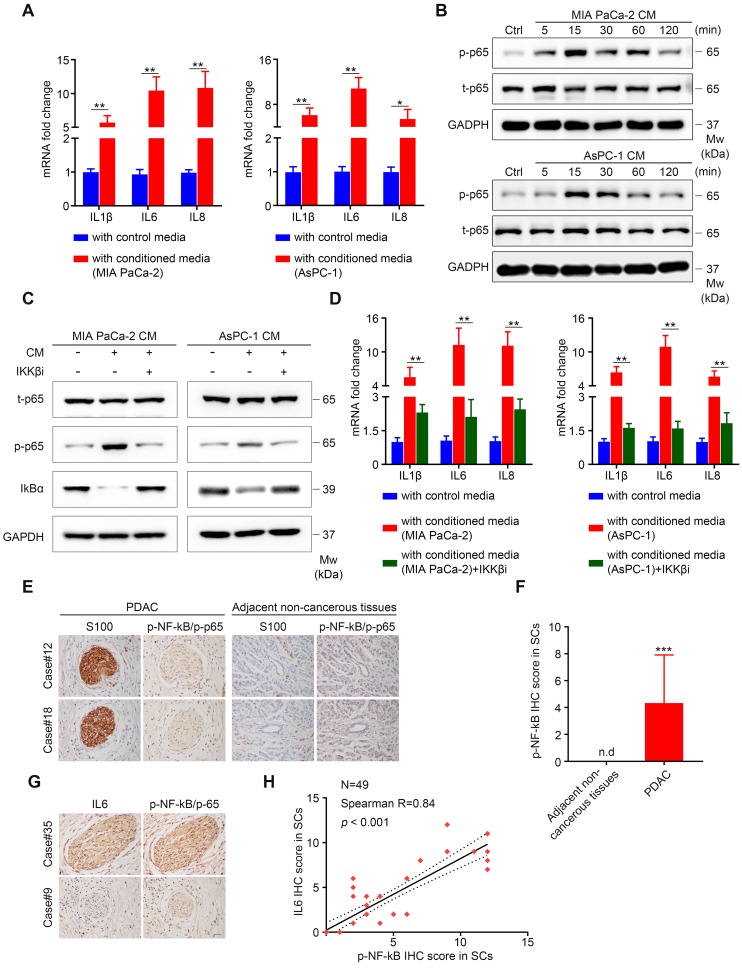
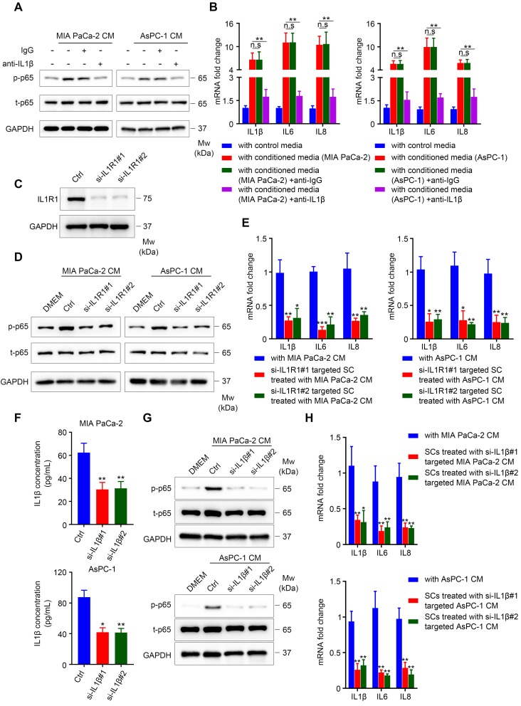
: The peripheral nervous system (PNS) plays an important role in tumor growth and progression. Schwann cells (SCs), the main glia cells of the PNS, augment cancer metastasis in contact-dependent or contact-independent manner in various malignancies. In the present study, we aimed to determine whether interplay between pancreatic cancer cells and SCs via paracrine signaling contributes to cancer progression. : Immunofluorescence analysis was performed to reveal the distribution of SCs in PDAC tissues and to determine the prognostic value and clinicopathological relevance of the level of intra‑tumoral SC markers for patients diagnosed with PDAC. Transwell assays and wound healing assays were carried out to investigate the influence of SC conditioned medium (SCM), SC co‑culture, or co-cultured CM on the migratory and invasive abilities of pancreatic cancer cells. The mechanism of SCs induced cancer cells migration and invasion was confirmed using quantitative real-time reverse transcription polymerase chain reaction (qRT-PCR), enzyme-linked immunosorbent assays (ELISAs), western blotting, immunofluorescence, immunohistochemistry, siRNA-mediated gene interference, and an mouse model. : Immunofluorescence analysis of tissue samples revealed that there were two different types of SCs distributed in the tumor microenvironment, the presence of which correlated with several clinicopathological characteristics and overall survival for patients with PDAC. Although SCM had no impact on the motility and invasiveness of tumor cells, both co-cultivation with SCs and co‑cultured CM enhanced pancreatic cancer cell migration and invasion. Mechanistically, SC‑derived Interleukin 6 (IL6), which was induced by co-culture with pancreatic cancer cells, augmented cancer cell migration and invasion by activating STAT3 signaling in cancer cells, while IL6 neutralization or STAT3 downregulation abrogated these effects. Furthermore, Interleukin 1β (IL1β), secreted by tumor cells, activated the nuclear actor (NF)-kappa B pathway in SCs, resulting in increased cytokines production, including IL6, while inhibiting the IL1β-IL1R1 axis led to inactivation of NF-kappa B signaling and downregulated cytokines expression in SCs. Interfering with tumor-neuroglia crosstalk impeded cancer cell dissemination . : Schwann cells were extensively distributed in the PDAC tumor microenvironment and high level of intra-tumoral SC markers could serve as an independent prognostic factor for poor survival of patients with PDAC. The tumor-neuroglia interaction is indispensable for SCs to acquire a tumor-facilitating phenotype. Targeting the tumor-neuroglia interplay might be a promising strategy to treat PDAC.
外周神经系统(PNS)在肿瘤生长和进展中起着重要作用。施万细胞(SCs)是 PNS 的主要神经胶质细胞,以依赖接触或非依赖接触的方式增强各种恶性肿瘤的癌症转移。在本研究中,我们旨在确定通过旁分泌信号的胰腺癌细胞与 SC 之间的相互作用是否有助于癌症进展。
免疫荧光分析用于揭示 SC 在 PDAC 组织中的分布,并确定 PDAC 患者肿瘤内 SC 标志物水平的预后价值和临床病理相关性。Transwell 测定和划痕愈合测定用于研究 SC 条件培养基(SCM)、SC 共培养或共培养 CM 对胰腺癌细胞迁移和侵袭能力的影响。使用定量实时逆转录聚合酶链反应(qRT-PCR)、酶联免疫吸附测定(ELISA)、Western blot、免疫荧光、免疫组织化学、siRNA 介导的基因干扰和小鼠模型证实了 SC 诱导癌细胞迁移和侵袭的机制。
组织样本的免疫荧光分析表明,存在两种分布在肿瘤微环境中的不同类型的 SC,其存在与 PDAC 患者的几种临床病理特征和总生存相关。虽然 SCM 对肿瘤细胞的运动性和侵袭性没有影响,但与 SC 的共培养和共培养 CM 均增强了胰腺癌细胞的迁移和侵袭。从机制上讲,由与胰腺癌细胞共培养诱导的 SC 衍生的白细胞介素 6(IL6)通过激活癌细胞中的 STAT3 信号增强了癌细胞的迁移和侵袭,而 IL6 中和或 STAT3 下调则消除了这些作用。此外,肿瘤细胞分泌的白细胞介素 1β(IL1β)激活了 SC 中的核因子(NF)-kappa B 途径,导致包括 IL6 在内的细胞因子产生增加,而抑制 IL1β-IL1R1 轴则导致 NF-kappa B 信号失活和 SC 中细胞因子表达下调。干扰肿瘤神经胶质细胞串扰会阻碍癌细胞的扩散。
施万细胞广泛分布在 PDAC 肿瘤微环境中,肿瘤内高水平的 SC 标志物可作为 PDAC 患者生存不良的独立预后因素。肿瘤神经胶质相互作用对于 SC 获得促进肿瘤的表型是必不可少的。靶向肿瘤神经胶质相互作用可能是治疗 PDAC 的一种有前途的策略。