• 文献检索
  • 文档翻译
  • 深度研究
  • 学术资讯
  • Suppr Zotero 插件Zotero 插件
  • 邀请有礼
  • 套餐&价格
  • 历史记录
应用&插件
Suppr Zotero 插件Zotero 插件浏览器插件Mac 客户端Windows 客户端微信小程序
定价
高级版会员购买积分包购买API积分包
服务
文献检索文档翻译深度研究API 文档MCP 服务
关于我们
关于 Suppr公司介绍联系我们用户协议隐私条款
关注我们

Suppr 超能文献

核心技术专利:CN118964589B侵权必究
粤ICP备2023148730 号-1Suppr @ 2026

文献检索

告别复杂PubMed语法,用中文像聊天一样搜索,搜遍4000万医学文献。AI智能推荐,让科研检索更轻松。

立即免费搜索

文件翻译

保留排版,准确专业,支持PDF/Word/PPT等文件格式,支持 12+语言互译。

免费翻译文档

深度研究

AI帮你快速写综述,25分钟生成高质量综述,智能提取关键信息,辅助科研写作。

立即免费体验

解析诱导多能干细胞来源的神经元发育过程中的蛋白质组动态变化。

Deciphering the Proteome Dynamics during Development of Neurons Derived from Induced Pluripotent Stem Cells.

机构信息

Biomolecular Mass Spectrometry and Proteomics, Bijvoet Center for Biomolecular Research and Utrecht Institute for Pharmaceutical Sciences, University of Utrecht, Padualaan 8, 3584 CH Utrecht, The Netherlands.

Netherlands Proteomics Center, Padualaan 8, 3584 CH Utrecht, The Netherlands.

出版信息

J Proteome Res. 2020 Jun 5;19(6):2391-2403. doi: 10.1021/acs.jproteome.0c00070. Epub 2020 May 15.

DOI:10.1021/acs.jproteome.0c00070
PMID:32357013
原文链接:https://pmc.ncbi.nlm.nih.gov/articles/PMC7281779/
Abstract

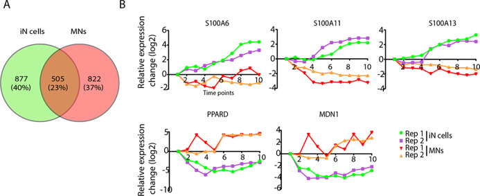
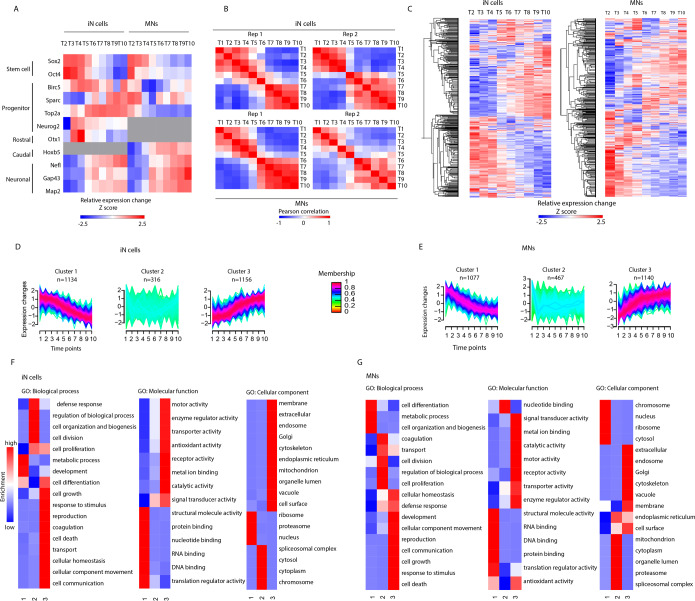
Neuronal development is a complex multistep process that shapes neurons by progressing though several typical stages, including axon outgrowth, dendrite formation, and synaptogenesis. Knowledge of the mechanisms of neuronal development is mostly derived from the study of animal models. Advances in stem cell technology now enable us to generate neurons from human induced pluripotent stem cells (iPSCs). Here we provide a mass spectrometry-based quantitative proteomic signature of human iPSC-derived neurons, i.e., iPSC-derived induced glutamatergic neurons and iPSC-derived motor neurons, throughout neuronal differentiation. Tandem mass tag 10-plex labeling was carried out to perform proteomic profiling of cells at different time points. Our analysis reveals significant expression changes (FDR < 0.001) of several key proteins during the differentiation process, e.g., proteins involved in the Wnt and Notch signaling pathways. Overall, our data provide a rich resource of information on protein expression during human iPSC neuron differentiation.

摘要

神经元发育是一个复杂的多步骤过程,通过经历几个典型阶段来塑造神经元,包括轴突生长、树突形成和突触发生。神经元发育机制的知识主要来自于动物模型的研究。干细胞技术的进步使我们能够从人类诱导多能干细胞 (iPSC) 中产生神经元。在这里,我们提供了基于质谱的人类 iPSC 衍生神经元的定量蛋白质组学特征,即 iPSC 衍生的兴奋性谷氨酸神经元和 iPSC 衍生的运动神经元,贯穿神经元分化过程。串联质量标签 10 plex 标记用于在不同时间点对细胞进行蛋白质组学分析。我们的分析揭示了分化过程中几个关键蛋白的显著表达变化 (FDR < 0.001),例如参与 Wnt 和 Notch 信号通路的蛋白。总的来说,我们的数据提供了人类 iPSC 神经元分化过程中蛋白质表达的丰富信息资源。

https://cdn.ncbi.nlm.nih.gov/pmc/blobs/b311/7281779/99fb15cb4107/pr0c00070_0005.jpg
https://cdn.ncbi.nlm.nih.gov/pmc/blobs/b311/7281779/d15881055b4a/pr0c00070_0001.jpg
https://cdn.ncbi.nlm.nih.gov/pmc/blobs/b311/7281779/808ca695d09e/pr0c00070_0002.jpg
https://cdn.ncbi.nlm.nih.gov/pmc/blobs/b311/7281779/8c03cf901d75/pr0c00070_0003.jpg
https://cdn.ncbi.nlm.nih.gov/pmc/blobs/b311/7281779/ec18381c3a78/pr0c00070_0004.jpg
https://cdn.ncbi.nlm.nih.gov/pmc/blobs/b311/7281779/99fb15cb4107/pr0c00070_0005.jpg

相似文献

1
Deciphering the Proteome Dynamics during Development of Neurons Derived from Induced Pluripotent Stem Cells.解析诱导多能干细胞来源的神经元发育过程中的蛋白质组动态变化。
J Proteome Res. 2020 Jun 5;19(6):2391-2403. doi: 10.1021/acs.jproteome.0c00070. Epub 2020 May 15.
2
Quantitative proteomic analysis of Rett iPSC-derived neuronal progenitors.基于 Rett 诱导多能干细胞的神经祖细胞的定量蛋白质组学分析。
Mol Autism. 2020 May 27;11(1):38. doi: 10.1186/s13229-020-00344-3.
3
Quantitative Map of Proteome Dynamics during Neuronal Differentiation.神经元分化过程中蛋白质组动力学的定量图谱
Cell Rep. 2017 Feb 7;18(6):1527-1542. doi: 10.1016/j.celrep.2017.01.025.
4
Quantitative profiling of axonal guidance proteins during the differentiation of human neurospheres.定量分析人神经球分化过程中轴突导向蛋白。
Biochim Biophys Acta Proteins Proteom. 2021 Aug;1869(8):140656. doi: 10.1016/j.bbapap.2021.140656. Epub 2021 Apr 20.
5
Generation and Proteome Profiling of PBMC-Originated, iPSC-Derived Corneal Endothelial Cells.原代单核细胞来源的诱导多能干细胞衍生的角膜内皮细胞的生成和蛋白质组分析。
Invest Ophthalmol Vis Sci. 2018 May 1;59(6):2437-2444. doi: 10.1167/iovs.17-22927.
6
Quantitative mapping of transcriptome and proteome dynamics during polarization of human iPSC-derived neurons.人诱导多能干细胞源性神经元极化过程中转录组和蛋白质组动态的定量图谱绘制。
Elife. 2020 Sep 17;9:e58124. doi: 10.7554/eLife.58124.
7
The proteomic architecture of schizophrenia iPSC-derived cerebral organoids reveals alterations in GWAS and neuronal development factors.精神分裂症诱导多能干细胞衍生的大脑类器官的蛋白质组结构揭示了全基因组关联研究和神经元发育因子的改变。
Transl Psychiatry. 2021 Oct 19;11(1):541. doi: 10.1038/s41398-021-01664-5.
8
Transcriptome and Proteome Profiling of Neural Induced Pluripotent Stem Cells from Individuals with Down Syndrome Disclose Dynamic Dysregulations of Key Pathways and Cellular Functions.唐氏综合征患者诱导多能干细胞的转录组和蛋白质组谱分析揭示了关键途径和细胞功能的动态失调。
Mol Neurobiol. 2019 Oct;56(10):7113-7127. doi: 10.1007/s12035-019-1585-3. Epub 2019 Apr 13.
9
Characterizing PCDH19 in human induced pluripotent stem cells (iPSCs) and iPSC-derived developing neurons: emerging role of a protein involved in controlling polarity during neurogenesis.在人类诱导多能干细胞(iPSC)和iPSC衍生的发育神经元中表征原钙黏蛋白19(PCDH19):一种参与神经发生过程中极性控制的蛋白质的新作用。
Oncotarget. 2015 Sep 29;6(29):26804-13. doi: 10.18632/oncotarget.5757.
10
Rho Kinase Inhibition Is Essential During In Vitro Neurogenesis and Promotes Phenotypic Rescue of Human Induced Pluripotent Stem Cell-Derived Neurons With Oligophrenin-1 Loss of Function.在体外神经发生过程中,Rho激酶抑制至关重要,并能促进具有少突神经胶质糖蛋白-1功能丧失的人诱导多能干细胞衍生神经元的表型拯救。
Stem Cells Transl Med. 2016 Jul;5(7):860-9. doi: 10.5966/sctm.2015-0303. Epub 2016 May 9.

引用本文的文献

1
Human sensory-like neuron surfaceome analysis.人类感觉样神经元表面蛋白质组分析。
PLoS One. 2025 Apr 2;20(4):e0320056. doi: 10.1371/journal.pone.0320056. eCollection 2025.
2
Proteomic Identification and Label-Free Quantification of Proteins Implicated in Neurite and Spine Formation.蛋白质组学鉴定和无标记定量分析参与神经突和脊柱形成的蛋白质。
Methods Mol Biol. 2024;2831:133-143. doi: 10.1007/978-1-0716-3969-6_10.
3
Single-cell transcriptome analysis reveals distinct cell populations in dorsal root ganglia and their potential roles in diabetic peripheral neuropathy.

本文引用的文献

1
Childhood-Onset Schizophrenia: Insights from Induced Pluripotent Stem Cells.儿童期发病精神分裂症:诱导多能干细胞的研究进展。
Int J Mol Sci. 2018 Nov 30;19(12):3829. doi: 10.3390/ijms19123829.
2
Distribution patterns of the zebrafish neuronal intermediate filaments inaa and inab.斑马鱼神经元中间丝 inaa 和 inab 的分布模式。
J Neurosci Res. 2019 Feb;97(2):202-214. doi: 10.1002/jnr.24347. Epub 2018 Nov 2.
3
Polymorphism Located in the Upstream Region of the Gene (rs2305809) Is Associated With Cervical Cancer: A Case-control Study.
单细胞转录组分析揭示了背根神经节中的不同细胞群体及其在糖尿病周围神经病变中的潜在作用。
PLoS One. 2024 Jul 31;19(7):e0306424. doi: 10.1371/journal.pone.0306424. eCollection 2024.
4
Expression Proteomics and Histone Analysis Reveal Extensive Chromatin Network Changes and a Role for Histone Tail Trimming during Cellular Differentiation.表达蛋白质组学和组蛋白分析揭示了广泛的染色质网络变化,以及组蛋白尾部修剪在细胞分化过程中的作用。
Biomolecules. 2024 Jun 24;14(7):747. doi: 10.3390/biom14070747.
5
Proteomics applications in next generation induced pluripotent stem cell models.蛋白质组学在新一代诱导多能干细胞模型中的应用。
Expert Rev Proteomics. 2024 Apr;21(4):217-228. doi: 10.1080/14789450.2024.2334033. Epub 2024 Mar 27.
6
Identification of MicroRNA Profiles in Fetal Spina Bifida: The Role in Pathomechanism and Diagnostic Significance.胎儿脊柱裂中微小RNA谱的鉴定:在发病机制中的作用及诊断意义
Int J Mol Sci. 2024 Mar 1;25(5):2896. doi: 10.3390/ijms25052896.
7
Multiomics Evaluation of Human iPSCs and iPSC-Derived Neurons.人类诱导多能干细胞及其衍生神经元的多组学评估
J Proteome Res. 2024 Aug 2;23(8):3149-3160. doi: 10.1021/acs.jproteome.3c00790. Epub 2024 Feb 28.
8
Macromolecular crowding in human tenocyte and skin fibroblast cultures: A comparative analysis.人肌腱细胞和皮肤成纤维细胞培养中的大分子拥挤:一项比较分析。
Mater Today Bio. 2024 Jan 28;25:100977. doi: 10.1016/j.mtbio.2024.100977. eCollection 2024 Apr.
9
Neurotoxic properties of the Zika virus envelope protein.寨卡病毒包膜蛋白的神经毒性。
Exp Neurol. 2023 Sep;367:114469. doi: 10.1016/j.expneurol.2023.114469. Epub 2023 Jun 14.
10
Integrated transcriptome and proteome analysis reveals posttranscriptional regulation of ribosomal genes in human brain organoids.整合转录组和蛋白质组分析揭示了人类脑类器官中核糖体基因的转录后调控。
Elife. 2023 Mar 29;12:e85135. doi: 10.7554/eLife.85135.
位于该基因上游区域的多态性(rs2305809)与宫颈癌相关:一项病例对照研究。
J Cancer Prev. 2018 Sep;23(3):147-152. doi: 10.15430/JCP.2018.23.3.147. Epub 2018 Sep 30.
4
Inhibition of collapsin response mediator protein-2 phosphorylation ameliorates motor phenotype of ALS model mice expressing SOD1G93A.抑制坍塌反应调节蛋白-2磷酸化可改善表达SOD1G93A的肌萎缩侧索硬化症模型小鼠的运动表型。
Neurosci Res. 2019 Feb;139:63-68. doi: 10.1016/j.neures.2018.08.016. Epub 2018 Sep 5.
5
Synaptic dysfunction in neurodegenerative and neurodevelopmental diseases: an overview of induced pluripotent stem-cell-based disease models.神经退行性和神经发育性疾病中的突触功能障碍:基于诱导多能干细胞的疾病模型概述。
Open Biol. 2018 Sep;8(9). doi: 10.1098/rsob.180138.
6
Current Advances and Limitations in Modeling ALS/FTD in a Dish Using Induced Pluripotent Stem Cells.利用诱导多能干细胞在培养皿中模拟肌萎缩侧索硬化症/额颞叶痴呆的研究进展与局限性
Front Neurosci. 2017 Dec 13;11:671. doi: 10.3389/fnins.2017.00671. eCollection 2017.
7
Pan-neuronal calcium imaging with cellular resolution in freely swimming zebrafish.在自由游动的斑马鱼中进行具有细胞分辨率的全神经元钙成像。
Nat Methods. 2017 Nov;14(11):1107-1114. doi: 10.1038/nmeth.4429. Epub 2017 Sep 11.
8
Gene expression profiles of brain endothelial cells during embryonic development at bulk and single-cell levels.胚胎发育过程中脑内皮细胞在整体水平和单细胞水平的基因表达谱。
Sci Signal. 2017 Jul 11;10(487):eaag2476. doi: 10.1126/scisignal.aag2476.
9
Spatiotemporal Proteomic Profiling of Human Cerebral Development.人类大脑发育的时空蛋白质组学分析
Mol Cell Proteomics. 2017 Sep;16(9):1548-1562. doi: 10.1074/mcp.M116.066274. Epub 2017 Jul 7.
10
Single-Cell RNA-Seq Reveals Hypothalamic Cell Diversity.单细胞RNA测序揭示下丘脑细胞多样性
Cell Rep. 2017 Mar 28;18(13):3227-3241. doi: 10.1016/j.celrep.2017.03.004.