Bingbing Gao, Yujing Zhou, Yanwei Miao, Chunbo Dong, Weiwei Wang, Shiyun Tian, Yangyingqiu Liu, Jin Shang, Qingwei Song, Ailian Liu, Lizhi Xie
Department of Radiology, First Affiliated Hospital of Dalian Medical University, Dalian, China.
GE Healthcare, MR Research, Beijing, China.
Front Neurol. 2020 Apr 17;11:252. doi: 10.3389/fneur.2020.00252. eCollection 2020.
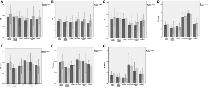
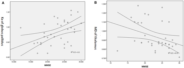
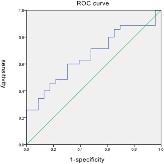
To explore the microstructural damage of extrapyramidal system gray matter nuclei in Parkinson disease (PD) using diffusion kurtosis imaging (DKI). We enrolled 35 clinically confirmed PD patients and 23 healthy volunteers. All patients underwent MR examination with conventional MRI scan sequences and an additional DKI sequence. We subsequently reconstructed the DKI raw images and analyzed the data. A radiologist in our hospital collected the Mini-Mental State Examination (MMSE) score of all subjects. In the PD group, the mean kurtosis and axial kurtosis level decreased in the red nucleus (RN) and thalamus; the radial kurtosis increased in the substantia nigra (SN) and globus pallidus (GP). Fractional anisotropy decreased in the putamen. The largest area under the ROC curve of mean diffusion in GP was 0.811. Most kurtosis parameters demonstrated a positive correlation with the MMSE score, while several diffusion parameters showed a negative correlation with the same. DKI can qualitatively distinguish PD from healthy controls; furthermore, DKI-derived parameters can quantitatively evaluate the modifications of microstructures in extrapyramidal system gray matter nucleus in PD.
利用扩散峰度成像(DKI)探讨帕金森病(PD)锥体外系灰质核团的微观结构损伤。我们纳入了35例临床确诊的PD患者和23名健康志愿者。所有患者均接受了常规MRI扫描序列及额外的DKI序列的MR检查。随后我们重建了DKI原始图像并分析数据。我院一名放射科医生收集了所有受试者的简易精神状态检查表(MMSE)评分。在PD组中,红核(RN)和丘脑的平均峰度及轴向峰度水平降低;黑质(SN)和苍白球(GP)的径向峰度增加。壳核的分数各向异性降低。GP平均扩散的ROC曲线下最大面积为0.811。大多数峰度参数与MMSE评分呈正相关,而一些扩散参数与MMSE评分呈负相关。DKI能够定性地区分PD患者与健康对照;此外,DKI衍生参数能够定量评估PD患者锥体外系灰质核团微观结构的改变。