Stanford University School of Medicine, Stanford, California.
Division of Allergy, Immunology, and Rheumatology, Department of Pediatrics, Stanford University School of Medicine, Stanford, California.
JAMA Netw Open. 2020 May 1;3(5):e204063. doi: 10.1001/jamanetworkopen.2020.4063.
Epidemiological studies indicate a link between obsessive-compulsive disorder and infections, particularly streptococcal pharyngitis. Pediatric acute-onset neuropsychiatric syndrome (PANS) manifests suddenly with obsessions, compulsions, and other behavioral disturbances, often after an infectious trigger. The current working model suggests a unifying inflammatory process involving the central nervous system, particularly the basal ganglia.
To investigate whether diffusion-weighted magnetic resonance imaging (DWI) detects microstructural abnormalities across the brain regions of children with PANS.
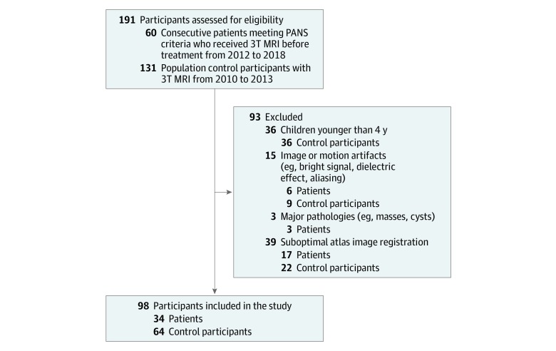
DESIGN, SETTING, AND PARTICIPANTS: Case-control study performed at a single-center, multidisciplinary clinic in the United States focusing on the evaluation and treatment of children with PANS. Sixty consecutive patients who underwent 3 Tesla (T) magnetic resonance imaging (MRI) before immunomodulation from September 3, 2012, to March 30, 2018, were retrospectively reviewed for study inclusion. Six patients were excluded by blinded investigators because of imaging or motion artifacts, 3 patients for major pathologies, and 17 patients for suboptimal atlas image registration. In total, 34 patients with PANS before initiation of treatment were compared with 64 pediatric control participants.
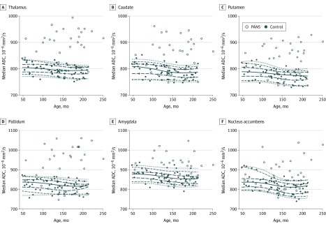
Using atlas-based MRI analysis, regional brain volume, diffusion, and cerebral blood flow were measured in the cerebral white matter, cerebral cortex, thalamus, caudate, putamen, pallidum, hippocampus, amygdala, nucleus accumbens, and brainstem. An age and sex-controlled multivariable analysis of covariance was used to compare patients with control participants.
This study compared 34 patients with PANS (median age, 154 months; age range, 55-251 months; 17 girls and 17 boys) and 64 pediatric control participants (median age, 139 months; age range, 48-213 months); 41 girls and 23 boys). Multivariable analysis demonstrated a statistically significant difference in MRI parameters between patients with PANS and control participants (F21,74 = 6.91; P < .001; partial η2 = 0.662). All assessed brain regions had statistically significantly increased median diffusivity compared with 64 control participants. Specifically, the deep gray matter (eg, the thalamus, basal ganglia, and amygdala) demonstrated the most profound increases in diffusivity consistent with the cardinal clinical symptoms of obsessions, compulsions, emotional dysregulation, and sleep disturbances. No statistically significant differences were found regarding volume and cerebral blood flow.
This study identifies cerebral microstructural differences in children with PANS in multiple brain structures, including the deep gray matter structures (eg, the thalamus, basal ganglia, and amygdala). Further study of MRI is warranted in prospective, clinical trials as a potential quantitative method for assessing patients under evaluation for PANS.
流行病学研究表明强迫症和感染之间存在关联,尤其是链球菌性咽炎。儿科急性发作的神经精神综合征(PANS)表现为突然出现的强迫观念、强迫行为和其他行为障碍,通常在感染触发后发生。目前的工作模型表明,涉及中枢神经系统的炎症过程具有统合性,特别是基底节。
研究弥散加权磁共振成像(DWI)是否能检测到患有 PANS 的儿童全脑区域的微观结构异常。
设计、地点和参与者: 这是一项在美国一家单中心多学科诊所进行的病例对照研究,该诊所专注于评估和治疗患有 PANS 的儿童。2012 年 9 月 3 日至 2018 年 3 月 30 日,对 60 名连续接受 3 特斯拉(T)磁共振成像(MRI)的患者进行了回顾性研究,以评估他们是否符合研究纳入标准。有 6 名患者被盲法研究者排除,原因是存在影像学或运动伪影、3 名患者存在主要病变,17 名患者的图谱图像配准效果欠佳。共比较了 34 名治疗前患有 PANS 的患者和 64 名儿科对照组患者。
使用基于图谱的 MRI 分析,在大脑白质、大脑皮层、丘脑、尾状核、壳核、苍白球、海马、杏仁核、伏隔核和脑干中测量了脑容量、扩散和脑血流。采用年龄和性别匹配的多元协方差分析比较患者和对照组。
这项研究比较了 34 名患有 PANS 的患者(中位数年龄 154 个月;年龄范围 55-251 个月;17 名女性和 17 名男性)和 64 名儿科对照组参与者(中位数年龄 139 个月;年龄范围 48-213 个月;41 名女性和 23 名男性)。多变量分析显示,PANS 患者和对照组患者的 MRI 参数存在统计学差异(F21,74=6.91;P<0.001;部分η2=0.662)。所有评估的脑区与 64 名对照组相比,中位扩散值均有统计学显著升高。具体而言,深部灰质(如丘脑、基底节和杏仁核)的扩散值增加最为显著,与强迫观念、强迫行为、情绪失调和睡眠障碍等主要临床症状一致。未发现体积和脑血流有统计学显著差异。
这项研究在多个脑结构中发现了患有 PANS 的儿童的脑微观结构差异,包括深部灰质结构(如丘脑、基底节和杏仁核)。需要进一步进行前瞻性临床试验的 MRI 研究,作为评估疑似 PANS 患者的潜在定量方法。