Institute of Neuropathology, University Medical Center Hamburg-Eppendorf (UKE), Hamburg, Germany.
Department of Neurology, Experimental Research in Stroke and Inflammation (ERSI), University Medical Center Hamburg-Eppendorf, Hamburg, Germany.
Mol Neurobiol. 2020 Jun;57(6):2812-2829. doi: 10.1007/s12035-020-01917-2. Epub 2020 May 4.
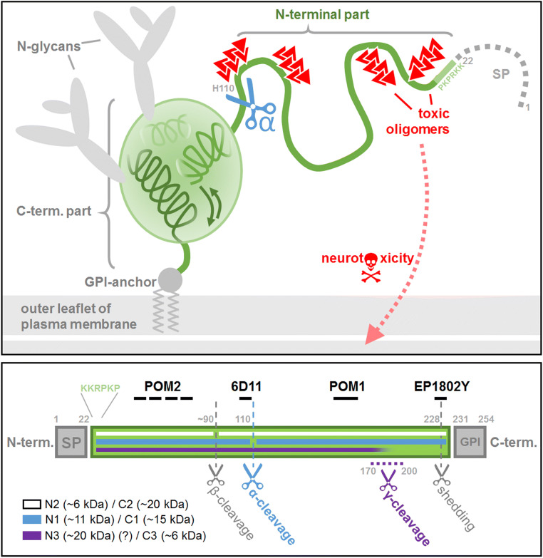
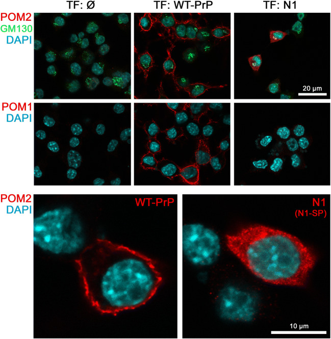
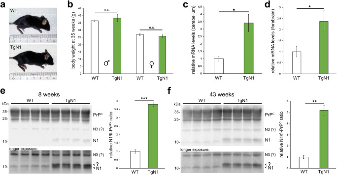
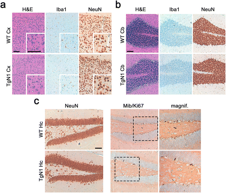
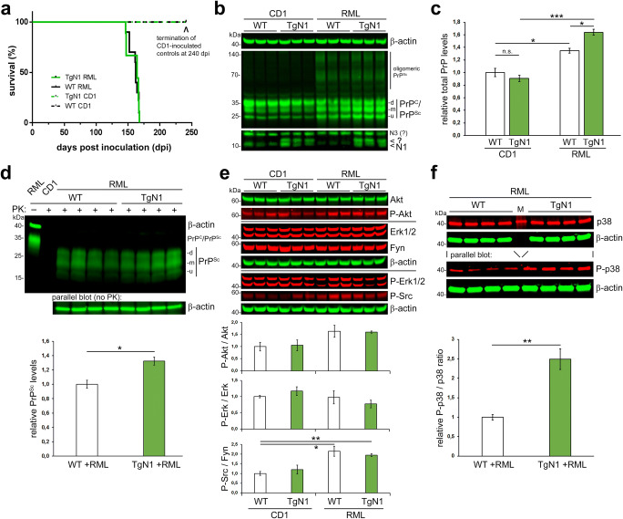
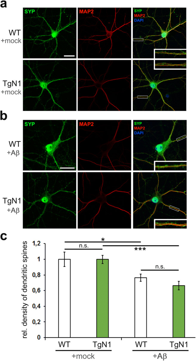
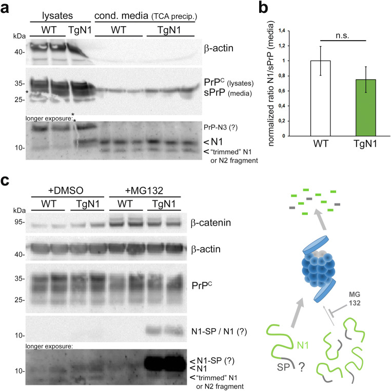
The structurally disordered N-terminal half of the prion protein (PrP) is constitutively released into the extracellular space by an endogenous proteolytic cleavage event. Once liberated, this N1 fragment acts neuroprotective in ischemic conditions and interferes with toxic peptides associated with neurodegenerative diseases, such as amyloid-beta (Aβ) in Alzheimer's disease. Since analog protective effects of N1 in prion diseases, such as Creutzfeldt-Jakob disease, have not been studied, and given that the protease releasing N1 has not been identified to date, we have generated and characterized transgenic mice overexpressing N1 (TgN1). Upon intracerebral inoculation of TgN1 mice with prions, no protective effects were observed at the levels of survival, clinical course, neuropathological, or molecular assessment. Likewise, primary neurons of these mice did not show protection against Aβ toxicity. Our biochemical and morphological analyses revealed that this lack of protective effects is seemingly due to an impaired ER translocation of the disordered N1 resulting in its cytosolic retention with an uncleaved signal peptide. Thus, TgN1 mice represent the first animal model to prove the inefficient ER translocation of intrinsically disordered domains (IDD). In contrast to earlier studies, our data challenge roles of cytoplasmic N1 as a cell penetrating peptide or as a potent "anti-prion" agent. Lastly, our study highlights both the importance of structured domains in the nascent chain for proteins to be translocated and aspects to be considered when devising novel N1-based therapeutic approaches against neurodegenerative diseases.
朊病毒蛋白(PrP)的结构无序 N 端前体通过内源性蛋白水解切割事件持续释放到细胞外空间。一旦释放,该 N1 片段在缺血条件下具有神经保护作用,并干扰与神经退行性疾病相关的毒性肽,如阿尔茨海默病中的淀粉样β(Aβ)。由于在朊病毒疾病(如克雅氏病)中尚未研究 N1 的类似保护作用,并且迄今为止尚未鉴定出释放 N1 的蛋白酶,因此我们生成并表征了过表达 N1(TgN1)的转基因小鼠。在将 TgN1 小鼠的脑中接种朊病毒后,在存活、临床病程、神经病理学或分子评估水平上均未观察到保护作用。同样,这些小鼠的原代神经元也未显示出对 Aβ 毒性的保护作用。我们的生化和形态分析表明,这种缺乏保护作用似乎是由于无序 N1 的内质网易位受损,导致其在细胞质中保留并带有未切割的信号肽。因此,TgN1 小鼠代表了首个证明内在无序域(ID)的内质网易位效率低下的动物模型。与早期的研究不同,我们的数据挑战了细胞质 N1 作为细胞穿透肽或作为有效“抗朊病毒”剂的作用。最后,我们的研究强调了新生链中结构域对于蛋白质易位的重要性,以及在设计针对神经退行性疾病的新型 N1 为基础的治疗方法时需要考虑的方面。