• 文献检索
  • 文档翻译
  • 深度研究
  • 学术资讯
  • Suppr Zotero 插件Zotero 插件
  • 邀请有礼
  • 套餐&价格
  • 历史记录
应用&插件
Suppr Zotero 插件Zotero 插件浏览器插件Mac 客户端Windows 客户端微信小程序
定价
高级版会员购买积分包购买API积分包
服务
文献检索文档翻译深度研究API 文档MCP 服务
关于我们
关于 Suppr公司介绍联系我们用户协议隐私条款
关注我们

Suppr 超能文献

核心技术专利:CN118964589B侵权必究
粤ICP备2023148730 号-1Suppr @ 2026

文献检索

告别复杂PubMed语法,用中文像聊天一样搜索,搜遍4000万医学文献。AI智能推荐,让科研检索更轻松。

立即免费搜索

文件翻译

保留排版,准确专业,支持PDF/Word/PPT等文件格式,支持 12+语言互译。

免费翻译文档

深度研究

AI帮你快速写综述,25分钟生成高质量综述,智能提取关键信息,辅助科研写作。

立即免费体验

氧化锌纳米颗粒诱导人诱导多能干细胞衍生心肌细胞中线粒体生物发生损伤和心脏功能障碍。

Zinc Oxide Nanoparticles Induce Mitochondrial Biogenesis Impairment and Cardiac Dysfunction in Human iPSC-Derived Cardiomyocytes.

机构信息

Graduate School, Academy of Military Medical Sciences, Beijing, People's Republic of China.

Department of Operational Medical Protection, PLA Center for Disease Control and Prevention, Beijing, People's Republic of China.

出版信息

Int J Nanomedicine. 2020 Apr 21;15:2669-2683. doi: 10.2147/IJN.S249912. eCollection 2020.

DOI:10.2147/IJN.S249912
PMID:32368048
原文链接:https://pmc.ncbi.nlm.nih.gov/articles/PMC7183345/
Abstract

BACKGROUND

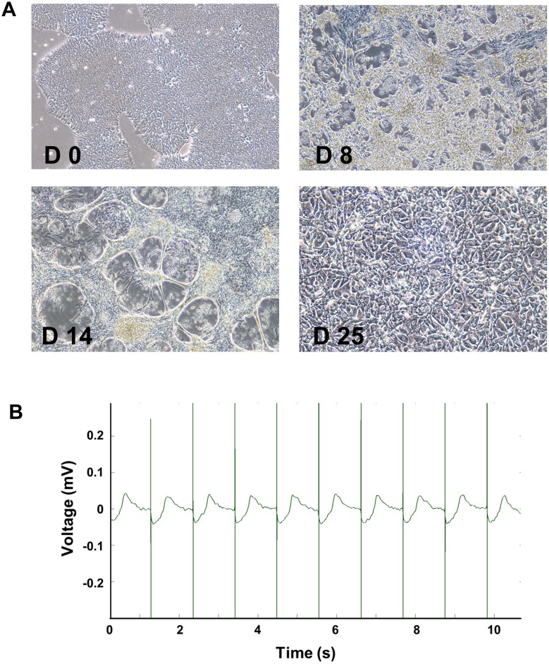
Zinc oxide nanoparticles (ZnO NPs) are one of the most widely used nanomaterials in a variety of fields such as industrial, pharmaceutical, and household applications. Increasing evidence suggests that ZnO NPs could elicit unignorable harmful effect to the cardiovascular system, but the potential deleterious effects to human cardiomyocytes remain to be elucidated. Human-induced pluripotent stem cell-derived cardiomyocytes (hiPSC-CMs) have been increasingly used as a promising in vitro model of cardiomyocyte in various fields such as drug cardiac safety evaluation. Herein, the present study was designed to elucidate the cardiac adverse effects of ZnO NPs and explore the possible underlying mechanism using hiPSC-CMs.

METHODS

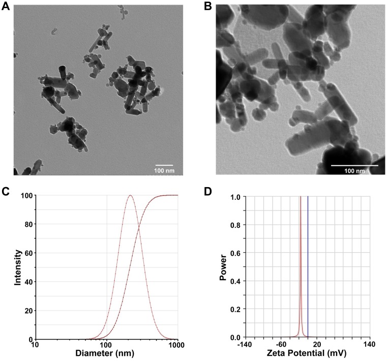
ZnO NPs were characterized by transmission electron microscopy and dynamic light scattering. The cytotoxicity induced by ZnO NPs in hiPSC-CMs was evaluated by determination of cell viability and lactate dehydrogenase release. Cellular reactive oxygen species (ROS) and mitochondrial membrane potential were measured by high-content analysis (HCA). Mitochondrial biogenesis was assayed by detection of mtDNA copy number and PGC-1α pathway. Moreover, microelectrode array techniques were used to investigate cardiac electrophysiological alterations.

RESULTS

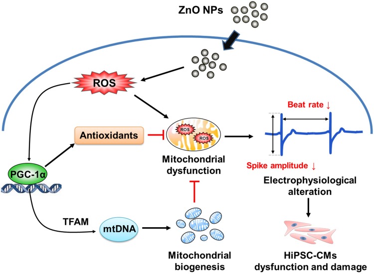
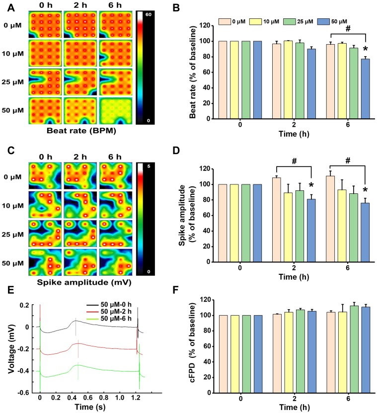
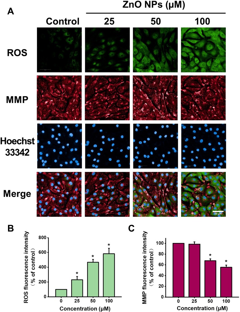
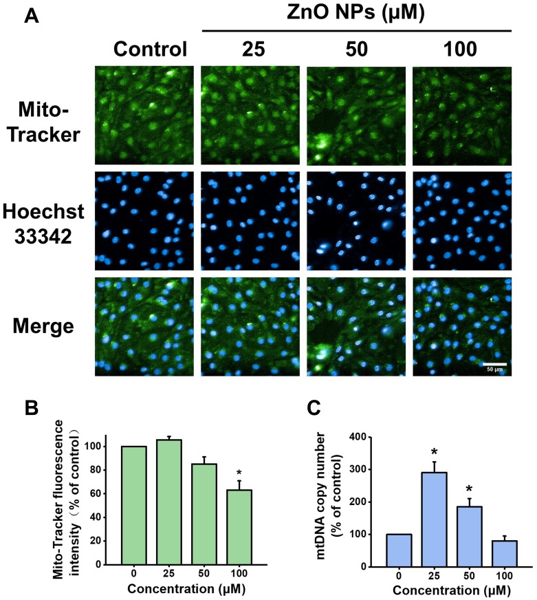
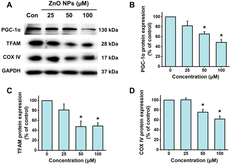
We demonstrated that ZnO NPs concentration- and time-dependently elicited cytotoxicity in hiPSC-CMs. The results from HCA revealed that ZnO NPs exposure at low-cytotoxic concentrations significantly promoted ROS generation and induced mitochondrial dysfunction. We further demonstrated that ZnO NPs could impair mitochondrial biogenesis and inhibit PGC-1α pathway. In addition, ZnO NPs at insignificantly cytotoxic concentrations were found to trigger cardiac electrophysiological alterations as evidenced by decreases of beat rate and spike amplitude.

CONCLUSION

Our findings unveiled the potential harmful effects of ZnO NPs to human cardiomyocytes that involve mitochondrial biogenesis and the PGC-1α pathway that could affect cardiac electrophysiological function.

摘要

背景

氧化锌纳米粒子(ZnO NPs)是一种在工业、制药和家庭应用等多种领域广泛应用的纳米材料。越来越多的证据表明,ZnO NPs 可能对心血管系统产生不可忽视的有害影响,但 ZnO NPs 对人类心肌细胞的潜在有害影响仍有待阐明。人诱导多能干细胞衍生的心肌细胞(hiPSC-CMs)已越来越多地被用作各种领域,如药物心脏安全性评估的心肌细胞的有前途的体外模型。在此,本研究旨在利用 hiPSC-CMs 阐明 ZnO NPs 的心脏不良影响,并探讨其潜在的机制。

方法

通过透射电子显微镜和动态光散射对 ZnO NPs 进行了表征。通过测定细胞活力和乳酸脱氢酶释放来评估 ZnO NPs 在 hiPSC-CMs 中的细胞毒性。通过高内涵分析(HCA)测定细胞内活性氧(ROS)和线粒体膜电位。通过检测 mtDNA 拷贝数和 PGC-1α 通路来测定线粒体生物发生。此外,还使用微电极阵列技术研究心脏电生理变化。

结果

我们证明了 ZnO NPs 浓度和时间依赖性地在 hiPSC-CMs 中产生细胞毒性。HCA 的结果表明,ZnO NPs 暴露在低细胞毒性浓度下会显著促进 ROS 生成并诱导线粒体功能障碍。我们进一步证明,ZnO NPs 可损害线粒体生物发生并抑制 PGC-1α 通路。此外,在无明显细胞毒性浓度下,ZnO NPs 被发现可触发心脏电生理变化,表现为心率和尖峰幅度降低。

结论

我们的研究结果揭示了 ZnO NPs 对人类心肌细胞的潜在有害影响,涉及线粒体生物发生和 PGC-1α 通路,可能影响心脏电生理功能。

https://cdn.ncbi.nlm.nih.gov/pmc/blobs/1e2b/7183345/bb5782482cbf/IJN-15-2669-g0008.jpg
https://cdn.ncbi.nlm.nih.gov/pmc/blobs/1e2b/7183345/d6ecff7cb6a9/IJN-15-2669-g0001.jpg
https://cdn.ncbi.nlm.nih.gov/pmc/blobs/1e2b/7183345/90e76cf9ee3a/IJN-15-2669-g0002.jpg
https://cdn.ncbi.nlm.nih.gov/pmc/blobs/1e2b/7183345/13adeed0e440/IJN-15-2669-g0003.jpg
https://cdn.ncbi.nlm.nih.gov/pmc/blobs/1e2b/7183345/b9e9c258d7c9/IJN-15-2669-g0004.jpg
https://cdn.ncbi.nlm.nih.gov/pmc/blobs/1e2b/7183345/6ab6369840b6/IJN-15-2669-g0005.jpg
https://cdn.ncbi.nlm.nih.gov/pmc/blobs/1e2b/7183345/ebe4a8cbe9b2/IJN-15-2669-g0006.jpg
https://cdn.ncbi.nlm.nih.gov/pmc/blobs/1e2b/7183345/a1c576966473/IJN-15-2669-g0007.jpg
https://cdn.ncbi.nlm.nih.gov/pmc/blobs/1e2b/7183345/bb5782482cbf/IJN-15-2669-g0008.jpg

相似文献

1
Zinc Oxide Nanoparticles Induce Mitochondrial Biogenesis Impairment and Cardiac Dysfunction in Human iPSC-Derived Cardiomyocytes.氧化锌纳米颗粒诱导人诱导多能干细胞衍生心肌细胞中线粒体生物发生损伤和心脏功能障碍。
Int J Nanomedicine. 2020 Apr 21;15:2669-2683. doi: 10.2147/IJN.S249912. eCollection 2020.
2
Bezafibrate Upregulates Mitochondrial Biogenesis and Influence Neural Differentiation of Human-Induced Pluripotent Stem Cells.贝扎贝特上调人诱导多能干细胞的线粒体生物发生并影响其神经分化。
Mol Neurobiol. 2019 Jun;56(6):4346-4363. doi: 10.1007/s12035-018-1368-2. Epub 2018 Oct 13.
3
TRPA1 promotes the maturation of embryonic stem cell-derived cardiomyocytes by regulating mitochondrial biogenesis and dynamics.TRPA1 通过调节线粒体生物发生和动力学促进胚胎干细胞来源的心肌细胞的成熟。
Stem Cell Res Ther. 2023 Jun 7;14(1):158. doi: 10.1186/s13287-023-03388-3.
4
Targeting HIF-1α in combination with PPARα activation and postnatal factors promotes the metabolic maturation of human induced pluripotent stem cell-derived cardiomyocytes.靶向 HIF-1α 与 PPARα 激活和出生后因素相结合可促进人诱导多能干细胞衍生心肌细胞的代谢成熟。
J Mol Cell Cardiol. 2019 Jul;132:120-135. doi: 10.1016/j.yjmcc.2019.05.003. Epub 2019 May 11.
5
Globular CTRP3 promotes mitochondrial biogenesis in cardiomyocytes through AMPK/PGC-1α pathway.球形 CTRP3 通过 AMPK/PGC-1α 通路促进心肌细胞中线粒体生物发生。
Biochim Biophys Acta Gen Subj. 2017 Jan;1861(1 Pt A):3085-3094. doi: 10.1016/j.bbagen.2016.10.022. Epub 2016 Oct 26.
6
NUCLEOLIN PROTECTS CARDIOMYOCYTES BY UPREGULATING PGC-1α AND PROMOTING MITOCHONDRIAL BIOGENESIS IN LPS-INDUCED MYOCARDIAL INJURY.在脂多糖诱导的心肌损伤中,核仁素通过上调PGC-1α和促进线粒体生物合成来保护心肌细胞。
Shock. 2023 Apr 1;59(4):627-636. doi: 10.1097/SHK.0000000000002084. Epub 2023 Jan 22.
7
Cytotoxicity of propofol in human induced pluripotent stem cell-derived cardiomyocytes.丙泊酚对人诱导多能干细胞来源心肌细胞的细胞毒性
J Anesth. 2018 Feb;32(1):120-131. doi: 10.1007/s00540-017-2441-0. Epub 2017 Dec 29.
8
NRF2 deficiency sensitizes human keratinocytes to zinc oxide nanoparticles-induced autophagy and cytotoxicity.NRF2 缺乏使人类角质形成细胞对氧化锌纳米颗粒诱导的自噬和细胞毒性敏感。
Environ Toxicol Pharmacol. 2021 Oct;87:103721. doi: 10.1016/j.etap.2021.103721. Epub 2021 Jul 30.
9
Overexpression of Peroxisome Proliferator-Activated Receptor γ Coactivator 1-α Protects Cardiomyocytes from Lipopolysaccharide-Induced Mitochondrial Damage and Apoptosis.过表达过氧化物酶体增殖物激活受体γ共激活因子 1-α 可保护心肌细胞免受脂多糖诱导的线粒体损伤和凋亡。
Inflammation. 2020 Oct;43(5):1806-1820. doi: 10.1007/s10753-020-01255-4.
10
PGC-1α promotes mitochondrial respiration and biogenesis during the differentiation of hiPSCs into cardiomyocytes.在人诱导多能干细胞分化为心肌细胞的过程中,PGC-1α促进线粒体呼吸和生物合成。
Genes Dis. 2020 Dec 23;8(6):891-906. doi: 10.1016/j.gendis.2020.12.006. eCollection 2021 Nov.

引用本文的文献

1
ZnO nanoparticles induce acute arrhythmia and heart failure in mice by disturbing cardiac ion channels.氧化锌纳米颗粒通过干扰心脏离子通道诱发小鼠急性心律失常和心力衰竭。
Front Cardiovasc Med. 2025 May 30;12:1569265. doi: 10.3389/fcvm.2025.1569265. eCollection 2025.
2
Long-Term Exposure of Fresh and Aged Nano Zinc Oxide Promotes Hepatocellular Carcinoma Malignancy by Up-Regulating Claudin-2.长期暴露于新鲜和老化纳米氧化锌通过上调 Claudin-2 促进肝癌恶性转化。
Int J Nanomedicine. 2024 Oct 1;19:9989-10008. doi: 10.2147/IJN.S478279. eCollection 2024.
3
Comprehensive insights into mechanism of nanotoxicity, assessment methods and regulatory challenges of nanomedicines.

本文引用的文献

1
hiPSCs Derived Cardiac Cells for Drug and Toxicity Screening and Disease Modeling: What Micro- Electrode-Array Analyses Can Tell Us.人多能干细胞衍生的心脏细胞在药物和毒性筛选及疾病建模中的应用:微电极阵列分析可以告诉我们什么。
Cells. 2019 Oct 28;8(11):1331. doi: 10.3390/cells8111331.
2
Subcellular Targets of Zinc Oxide Nanoparticles During the Aging Process: Role of Cross-talk Between Mitochondrial Dysfunction and Endoplasmic Reticulum Stress in the Genotoxic Response.衰老过程中氧化锌纳米颗粒的亚细胞靶点:线粒体功能障碍与内质网应激之间的相互作用在遗传毒性反应中的作用
Toxicol Sci. 2019 Sep 1;171(1):159-171. doi: 10.1093/toxsci/kfz132.
3
对纳米毒性机制、纳米药物评估方法及监管挑战的全面洞察。
Discov Nano. 2024 Oct 4;19(1):165. doi: 10.1186/s11671-024-04118-1.
4
Electrophysiological and biochemical effect of zinc oxide nanoparticles on heart functions of male Wistar rats.氧化锌纳米颗粒对雄性 Wistar 大鼠心脏功能的电生理和生化效应。
Sci Rep. 2024 Jul 4;14(1):15416. doi: 10.1038/s41598-024-65189-9.
5
On the Role of Starchy Grains in Ice Nucleation Processes.淀粉颗粒在冰核形成过程中的作用
ACS Food Sci Technol. 2024 Apr 11;4(5):1039-1051. doi: 10.1021/acsfoodscitech.3c00561. eCollection 2024 May 17.
6
Biogenic Zinc Oxide Nanoparticles synthesized from induce oxidative stress, mitochondrial damage and apoptosis in Colorectal Cancer.由 合成的生物成因氧化锌纳米粒子可诱导结直肠癌中的氧化应激、线粒体损伤和细胞凋亡。
Nanotheranostics. 2024 Mar 9;8(3):312-329. doi: 10.7150/ntno.84995. eCollection 2024.
7
Impact of peripheral blood mononuclear cells preconditioned by activated platelet supernatant in managing gastric mucosal damage induced by zinc oxide nanoparticles in rats.活化血小板上清液预处理的外周血单个核细胞对大鼠氧化锌纳米颗粒诱导的胃黏膜损伤的影响
Anat Cell Biol. 2024 Mar 31;57(1):105-118. doi: 10.5115/acb.23.217. Epub 2024 Jan 15.
8
Protective Role of Short-term Aerobic Exercise Against Zinc Oxide Nanoparticles-Induced Cardiac Oxidative Stress Via Possible Changes of Apelin, Angiotensin II/Angiotensin II Type I Signalling Pathway.短期有氧运动通过可能改变的 Apelin、血管紧张素 II/血管紧张素 II 型信号通路对氧化锌纳米颗粒诱导的心脏氧化应激的保护作用。
Cardiovasc Toxicol. 2023 Jun;23(5-6):177-184. doi: 10.1007/s12012-023-09792-8. Epub 2023 May 15.
9
Experimental Methods for the Biological Evaluation of Nanoparticle-Based Drug Delivery Risks.基于纳米颗粒的药物递送风险生物评估的实验方法
Pharmaceutics. 2023 Feb 11;15(2):612. doi: 10.3390/pharmaceutics15020612.
10
Zinc Oxide Nanoparticles and Synthesized Pyrazolopyrimidine Alleviate Diabetic Effects in Rats Induced by Type II Diabetes.氧化锌纳米颗粒与合成的吡唑并嘧啶减轻II型糖尿病诱导的大鼠糖尿病效应。
ACS Omega. 2022 Oct 6;7(41):36865-36872. doi: 10.1021/acsomega.2c05638. eCollection 2022 Oct 18.
Doxorubicin-induced cardiotoxicity is maturation dependent due to the shift from topoisomerase IIα to IIβ in human stem cell derived cardiomyocytes.
多柔比星诱导的心脏毒性与成熟度相关,这是由于人源干细胞来源的心肌细胞中拓扑异构酶 IIα 向 IIβ 的转变所致。
J Cell Mol Med. 2019 Jul;23(7):4627-4639. doi: 10.1111/jcmm.14346. Epub 2019 May 20.
4
Mitochondrial proteins: from biogenesis to functional networks.线粒体蛋白:从生物发生到功能网络。
Nat Rev Mol Cell Biol. 2019 May;20(5):267-284. doi: 10.1038/s41580-018-0092-0.
5
International Multisite Study of Human-Induced Pluripotent Stem Cell-Derived Cardiomyocytes for Drug Proarrhythmic Potential Assessment.国际多中心人类诱导多能干细胞衍生心肌细胞药物致心律失常潜能评估研究。
Cell Rep. 2018 Sep 25;24(13):3582-3592. doi: 10.1016/j.celrep.2018.08.079.
6
Zinc oxide nanoparticles induce toxicity by affecting cell wall integrity pathway, mitochondrial function and lipid homeostasis in Saccharomyces cerevisiae.氧化锌纳米颗粒通过影响酿酒酵母细胞壁完整性途径、线粒体功能和脂质动态平衡诱导毒性。
Chemosphere. 2018 Dec;213:65-75. doi: 10.1016/j.chemosphere.2018.09.028. Epub 2018 Sep 5.
7
A review of cardiovascular toxicity of TiO, ZnO and Ag nanoparticles (NPs).纳米 TiO₂、ZnO 和 Ag 纳米粒子的心血管毒性综述。
Biometals. 2018 Aug;31(4):457-476. doi: 10.1007/s10534-018-0113-7. Epub 2018 May 10.
8
Doxorubicin-induced mitophagy and mitochondrial damage is associated with dysregulation of the PINK1/parkin pathway.多柔比星诱导的线粒体自噬和线粒体损伤与 PINK1/parkin 通路的失调有关。
Toxicol In Vitro. 2018 Sep;51:1-10. doi: 10.1016/j.tiv.2018.05.001. Epub 2018 May 3.
9
Cross-Site Reliability of Human Induced Pluripotent stem cell-derived Cardiomyocyte Based Safety Assays Using Microelectrode Arrays: Results from a Blinded CiPA Pilot Study.基于微电极阵列的人诱导多能干细胞源性心肌细胞安全性检测的跨站点可靠性:盲法 CiPA 初步研究结果。
Toxicol Sci. 2018 Aug 1;164(2):550-562. doi: 10.1093/toxsci/kfy110.
10
Zinc oxide nanoparticles induce HIF-1α protein stabilization through increased reactive oxygen species generation from electron transfer chain complex III of mitochondria.氧化锌纳米颗粒通过增加线粒体电子传递链复合物III产生的活性氧来诱导缺氧诱导因子-1α蛋白稳定。
J Dermatol Sci. 2018 Jul;91(1):104-107. doi: 10.1016/j.jdermsci.2018.03.010. Epub 2018 Mar 20.