Shen Feng, Pan Xiangou, Li Min, Chen Yixing, Jiang Ying, He Jian
Department of Medical Oncology, Zhongshan Hospital, Fudan University, Shanghai 200032, People's Republic of China.
Department of Radiation Oncology, Zhongshan Hospital, Fudan University, Shanghai 200032, People's Republic of China.
Onco Targets Ther. 2020 Apr 16;13:3165-3176. doi: 10.2147/OTT.S246899. eCollection 2020.
Breast cancer remains a great threat to females worldwide. As a recently defined programmed cell death pathway that associates with immune activation, RIP1/RIP3/MLKL necroptosis signaling has been implicated in a variety of diseases. The present study aimed to investigate the role of RIP1/RIP3/MLKL signaling in breast cancer cell proliferation and metastasis in vivo and in vitro.
Western blot and quantitative real-time PCR were performed to evaluate the activation of necroptosis signaling in clinical human breast cancer tissues. Correlation of necroptosis signaling markers with clinicopathological parameters was statistically assessed. Cell viability assay, colony formation assay, wound healing assay, and transwell migration and invasion assays were performed to investigate the effects of necroptosis inhibition on breast cancer cell proliferation and metastasis.
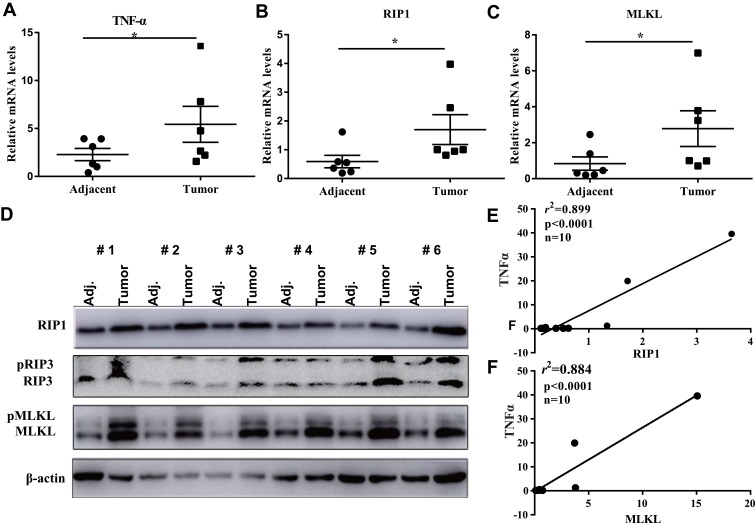
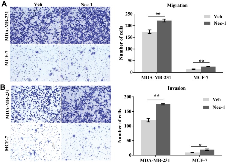
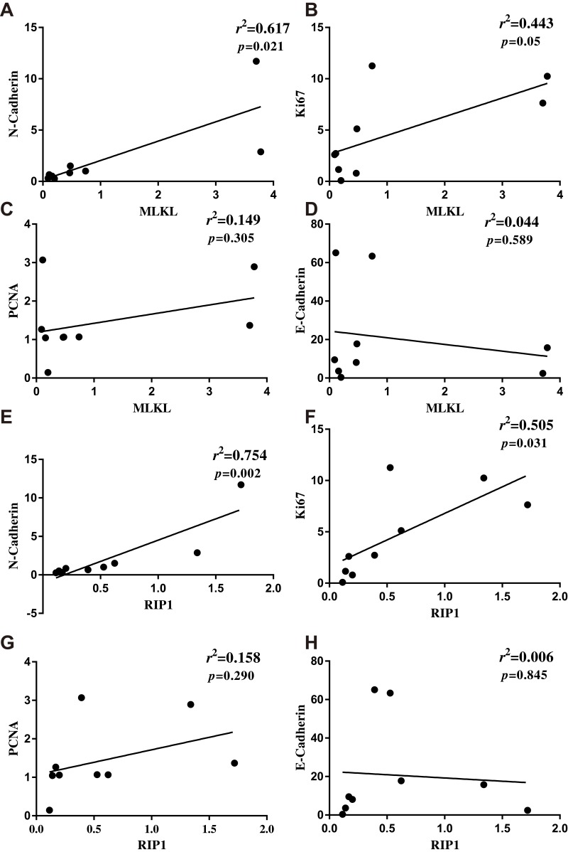
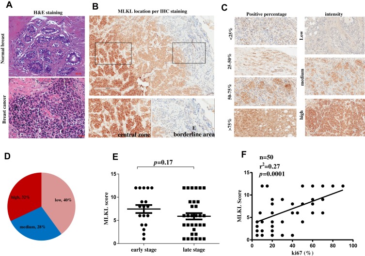
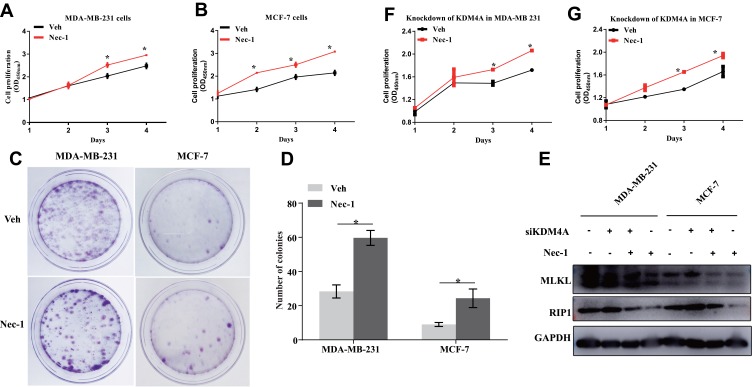
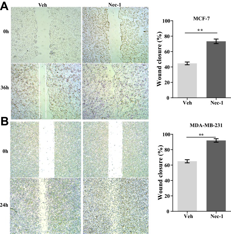
Clinical breast cancer tissues showed significantly higher levels of tumor necrosis factor alpha (TNFα), RIP1, RIP3 and MLKL at both mRNA and protein levels as compared with their paired non-cancerous tissues. Phosphorylation of RIP3 and MLKL was also remarkably provoked. Statistics showed that both RIP1 and MLKL positively correlated with cancer parameters such as N-cadherin (=0.002 for RIP1 and =0.021 for MLKL) and Ki67 (=0.031 for RIP1 and =0.05 for MLKL). The MLKL expression level significantly correlated with tumor size (=0.001) and the proliferation indicator Ki67 (=0.018). In addition, pharmacological inhibition of the necroptosis signaling using necrostatin-1 promoted breast cancer cell proliferation and colony formation by approximately 50%. Blockade of necroptosis signaling also accelerated wound healing process and cell transmigration in breast cancer cells.
Our results suggested that pharmacological inhibition of necroptosis promoted breast cancer cell proliferation and metastasis. Modulation of tumor cell necroptosis might represent a novel strategy as to breast cancer treatment.
乳腺癌仍然是全球女性面临的重大威胁。作为一种最近定义的与免疫激活相关的程序性细胞死亡途径,RIP1/RIP3/MLKL坏死性凋亡信号通路已涉及多种疾病。本研究旨在探讨RIP1/RIP3/MLKL信号通路在体内外乳腺癌细胞增殖和转移中的作用。
采用蛋白质免疫印迹法和定量实时聚合酶链反应评估临床人乳腺癌组织中坏死性凋亡信号通路的激活情况。对坏死性凋亡信号标志物与临床病理参数的相关性进行统计学评估。进行细胞活力测定、集落形成测定、伤口愈合测定以及Transwell迁移和侵袭测定,以研究坏死性凋亡抑制对乳腺癌细胞增殖和转移的影响。
与配对的癌旁组织相比,临床乳腺癌组织在mRNA和蛋白质水平上肿瘤坏死因子α(TNFα)、RIP1、RIP3和MLKL的表达水平均显著升高。RIP3和MLKL的磷酸化也明显增强。统计学分析表明,RIP1和MLKL均与癌症参数如N-钙黏蛋白(RIP1为=0.002,MLKL为=0.021)和Ki67(RIP1为=0.031,MLKL为=0.05)呈正相关。MLKL表达水平与肿瘤大小(=0.001)和增殖指标Ki67(=0.018)显著相关。此外,使用坏死性凋亡抑制剂necrostatin-1对坏死性凋亡信号通路进行药理学抑制可使乳腺癌细胞增殖和集落形成增加约50%。阻断坏死性凋亡信号通路还可加速乳腺癌细胞的伤口愈合过程和细胞迁移。
我们的结果表明,对坏死性凋亡进行药理学抑制可促进乳腺癌细胞的增殖和转移。调节肿瘤细胞坏死性凋亡可能是一种新的乳腺癌治疗策略。