Department of Clinical and Experimental Epilepsy, University College London Queen Square Institute of Neurology, London, United Kingdom.
MRI Unit, Epilepsy Society, Chalfont St Peter, United Kingdom.
Ann Neurol. 2020 Jul;88(1):170-182. doi: 10.1002/ana.25762. Epub 2020 May 28.
Cognitive problems, especially disturbances in episodic memory, and hippocampal sclerosis are common in temporal lobe epilepsy (TLE), but little is known about the relationship of hippocampal morphology with memory. We aimed to relate hippocampal surface-shape patterns to verbal and visual learning.
We analyzed hippocampal surface shapes on high-resolution magnetic resonance images and the Adult Memory and Information Processing Battery in 145 unilateral refractory TLE patients undergoing epilepsy surgery, a validation set of 55 unilateral refractory TLE patients, and 39 age- and sex-matched healthy volunteers.
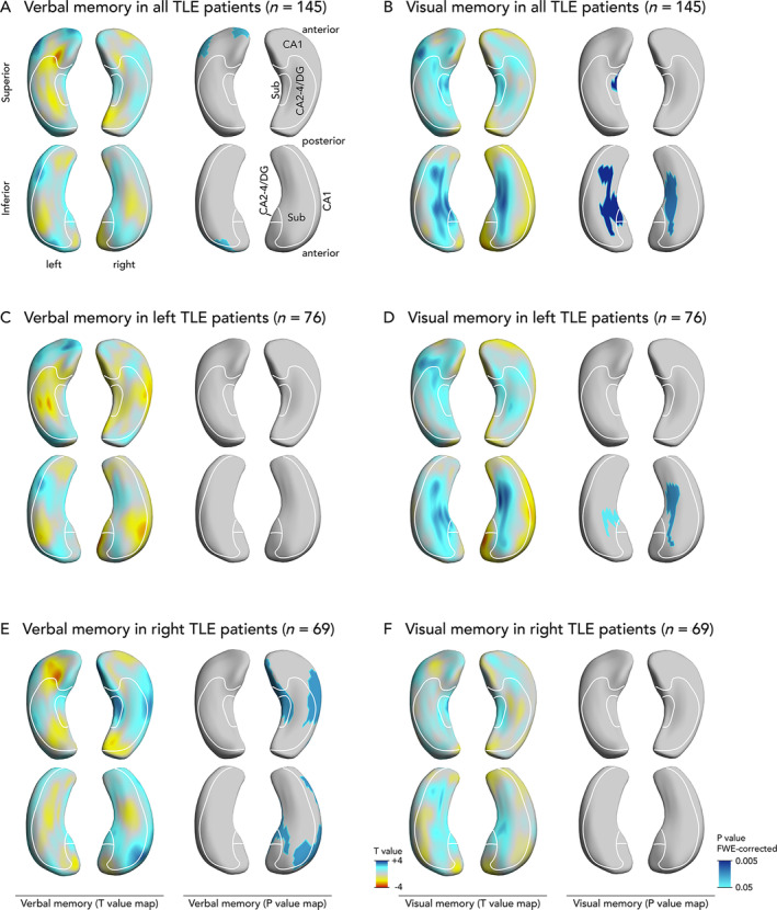
Both left TLE (LTLE) and right TLE (RTLE) patients had lower verbal (LTLE 44 ± 11; RTLE 45 ± 10) and visual learning (LTLE 34 ± 8, RTLE 30 ± 8) scores than healthy controls (verbal 58 ± 8, visual 39 ± 6; p < 0.001). Verbal learning was more impaired the greater the atrophy of the left superolateral hippocampal head. In contrast, visual memory was worse with greater bilateral inferomedial hippocampal atrophy. Postsurgical verbal memory decline was more common in LTLE than in RTLE (reliable change index in LTLE 27% vs RTLE 7%, p = 0.006), whereas there were no differences in postsurgical visual memory decline between those groups. Preoperative atrophy of the left hippocampal tail predicted postsurgical verbal memory decline.
Memory deficits in TLE are associated with specific morphological alterations of the hippocampus, which could help stratify TLE patients into those at high versus low risk of presurgical or postsurgical memory deficits. This knowledge could improve planning and prognosis of selective epilepsy surgery and neuropsychological counseling in TLE. ANN NEUROL 2020 ANN NEUROL 2020;88:170-182.
认知问题,尤其是情景记忆障碍,以及海马硬化,在颞叶癫痫(TLE)中很常见,但对于海马形态与记忆的关系知之甚少。我们旨在研究海马表面形态与言语和视觉学习的关系。
我们分析了 145 例接受癫痫手术的单侧难治性 TLE 患者、55 例单侧难治性 TLE 患者验证集和 39 名年龄和性别匹配的健康志愿者的高分辨率磁共振图像和成人记忆和信息处理电池的海马表面形状。
左 TLE(LTLE)和右 TLE(RTLE)患者的言语(LTLE 44 ± 11;RTLE 45 ± 10)和视觉学习(LTLE 34 ± 8,RTLE 30 ± 8)得分均低于健康对照组(言语 58 ± 8,视觉 39 ± 6;p < 0.001)。左海马头外侧萎缩越严重,言语学习障碍越严重。相反,双侧海马内下萎缩越严重,视觉记忆越差。LTLE 术后言语记忆下降比 RTLE 更常见(LTLE 的可靠变化指数为 27%,RTLE 为 7%,p = 0.006),而两组间术后视觉记忆下降无差异。术前左海马尾部萎缩预测术后言语记忆下降。
TLE 的记忆缺陷与海马的特定形态改变有关,这有助于将 TLE 患者分为术前或术后记忆缺陷风险高与低的患者。这些知识可以改善选择性癫痫手术的规划和预后,并为 TLE 提供神经心理学咨询。