Zawar Ifrah, Zhu Shen, Quigg Mark, Kapur Jaideep, Manning Carol, Fletcher P Thomas
Comprehensive Epilepsy Program, Department of Neurology, University of Virginia, Charlottesville, VA 22908, USA.
Department of Computer Science, University of Virginia, Charlottesville, VA 22908, USA.
Neuroimage Clin. 2025 Jun 18;47:103830. doi: 10.1016/j.nicl.2025.103830.
Epilepsy is common in Alzheimer's disease (AD) and non-AD dementias. However, the neuroimaging correlates of epilepsy in dementias remain unexplored. We investigated mesial temporal morphology and volumes in AD (AD + Epi) and nonAD dementias (nonAD + Epi) with epilepsy.
Participants from 39 US Alzheimer's disease centers (9/2005-12/2021) were classified into dementia with epilepsy (AD + Epi, nonAD + Epi), dementia without epilepsy (AD-Epi, nonAD-Epi); and healthy controls. Dementia with epilepsy participants with available MRIs were matched to dementia without epilepsy and healthy controls by age, sex, and dementia type (AD versus non-AD). FreeSurfer segmented hippocampi and amygdalae. Point distribution models created via ShapeWorks quantified morphological differences in the left and right hippocampi and amygdalae. Hippocampal and amygdalar volumes were normalized to the total intracranial volume. Multivariate analysis of covariates (MANCOVA), adjusted for age, sex, intracranial volume, and dementia severity, identified statistically significant local morphological and normalized volume group differences.
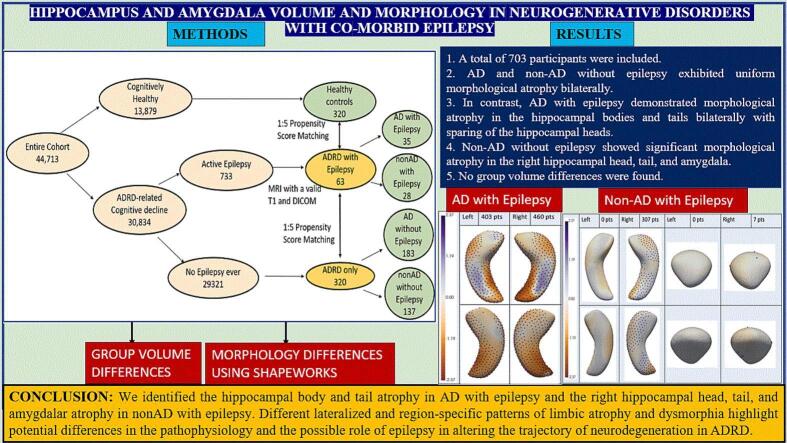
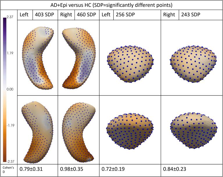
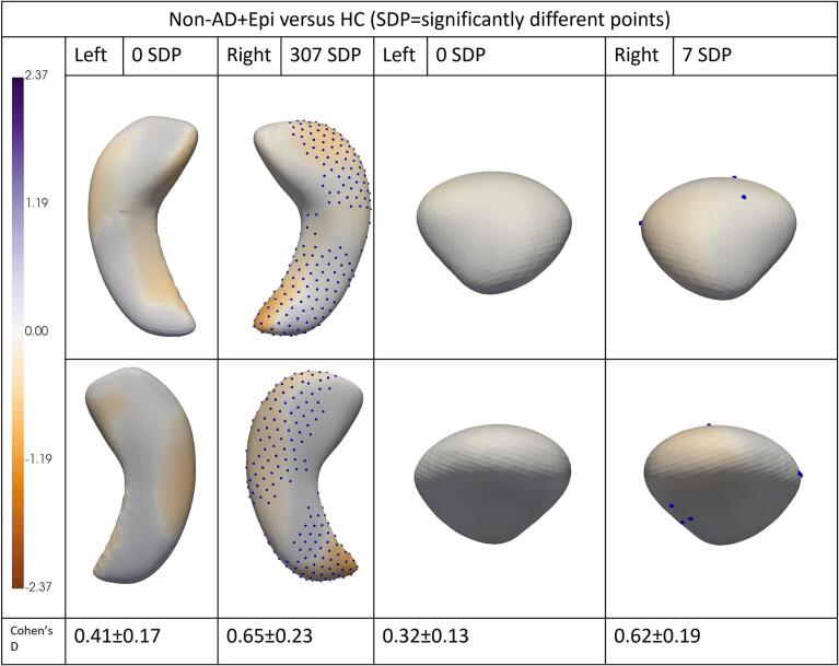
A total of 703 participants (average age: 70.78 years, 391 (55.62 %) female) were included. AD-Epi and NonAD-Epi exhibited uniform hippocampal and amygdalar morphological atrophy bilaterally. In contrast, AD + Epi demonstrated morphological atrophy in the hippocampal bodies and tails bilaterally with sparing of the hippocampal heads, more pronounced inward deviations on mesial and lateral surfaces, and outward deviations in the middle hippocampal body bilaterally on the superior surface. NonAD + Epi showed significant morphological atrophy in the right hippocampal head, tail, and amygdala. No group volume differences were found.
We identified hippocampal body and tail atrophy in AD + Epi and right hippocampal head, tail, and amygdalar atrophy in nonAD + Epi. Different lateralized and region-specific patterns of limbic atrophy and dysmorphia highlight potential differences in the pathophysiology and the possible role of epilepsy in altering the trajectory of neurodegeneration in AD and nonAD.
癫痫在阿尔茨海默病(AD)和非AD痴呆中很常见。然而,痴呆中癫痫的神经影像学相关性仍未得到探索。我们研究了患有癫痫的AD(AD + Epi)和非AD痴呆(非AD + Epi)的内侧颞叶形态和体积。
来自美国39个阿尔茨海默病中心(2005年9月 - 2021年12月)的参与者被分为伴有癫痫的痴呆(AD + Epi,非AD + Epi)、不伴有癫痫的痴呆(AD - Epi,非AD - Epi)以及健康对照。对有可用MRI的伴有癫痫的痴呆参与者,按年龄、性别和痴呆类型(AD与非AD)与不伴有癫痫的痴呆和健康对照进行匹配。FreeSurfer软件分割海马体和杏仁核。通过ShapeWorks创建的点分布模型量化左右海马体和杏仁核的形态差异。将海马体和杏仁核体积标准化为总颅内体积。对年龄、性别、颅内体积和痴呆严重程度进行调整的协变量多变量分析(MANCOVA)确定了具有统计学意义的局部形态和标准化体积组间差异。
共纳入703名参与者(平均年龄:70.78岁,391名(55.62%)为女性)。AD - Epi和非AD - Epi双侧海马体和杏仁核均表现出均匀的形态萎缩。相比之下,AD + Epi双侧海马体体部和尾部出现形态萎缩,海马头部未受累,内侧和外侧表面向内偏移更明显,上表面双侧海马体中部向外偏移。非AD + Epi右侧海马头部、尾部和杏仁核出现明显的形态萎缩。未发现组间体积差异。
我们发现AD + Epi中海马体体部和尾部萎缩,非AD + Epi中右侧海马头部、尾部和杏仁核萎缩。不同的侧化和区域特异性边缘系统萎缩及畸形模式突出了AD和非AD中病理生理学的潜在差异以及癫痫在改变神经退行性变轨迹中可能发挥的作用。