Institut de la Vision, Sorbonne Université, INSERM, CNRS, Paris, France.
Centre Hospitalier National d'Ophtalmologie des 15-20, DHU Sight Restore, INSERM-DHOS CIC, Paris, France.
Stem Cells Transl Med. 2020 Aug;9(8):917-935. doi: 10.1002/sctm.19-0306. Epub 2020 May 7.
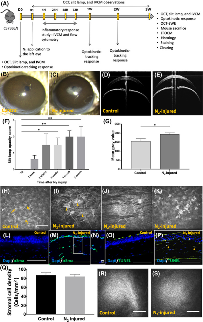
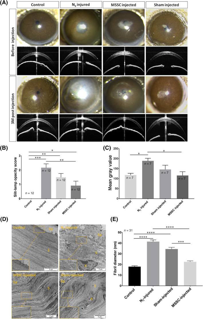
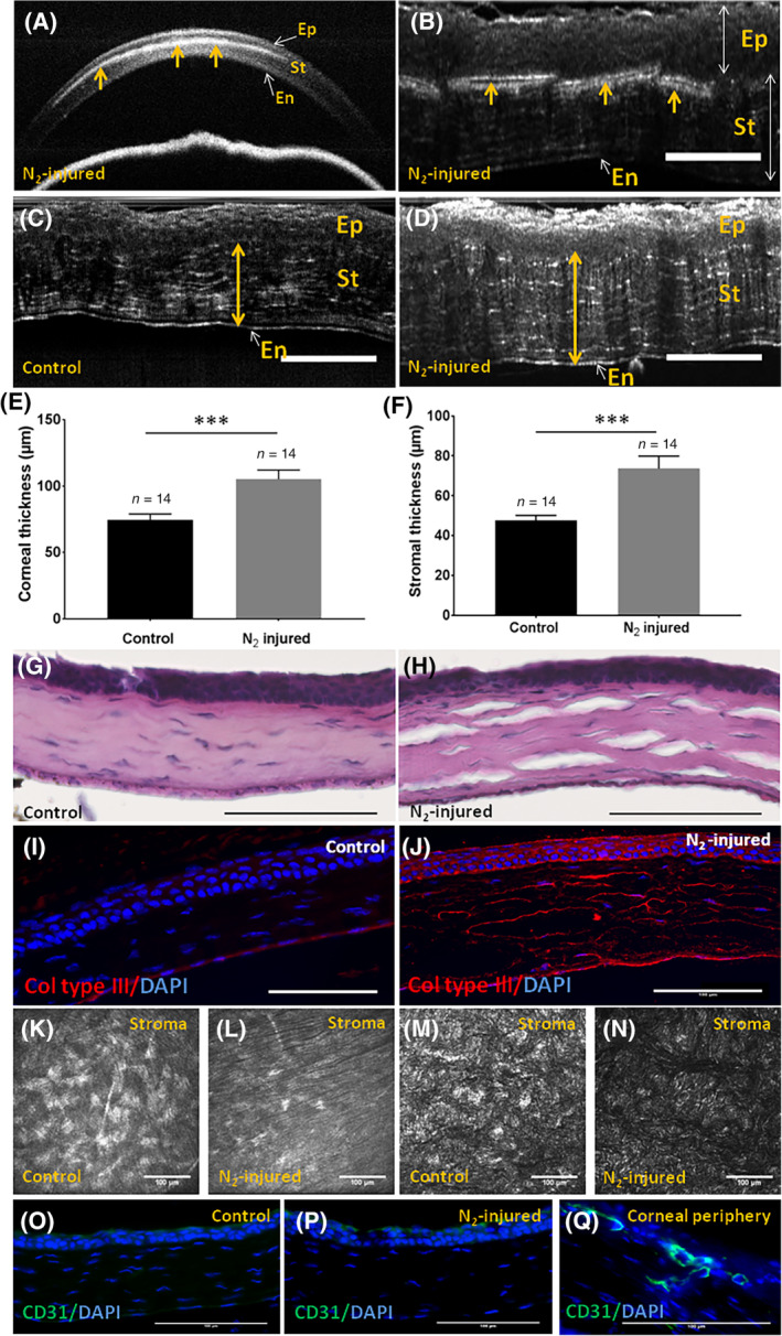
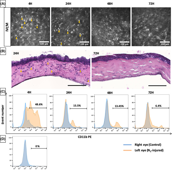
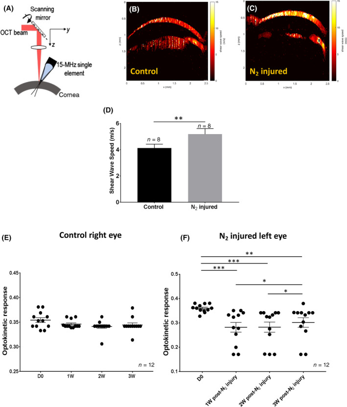
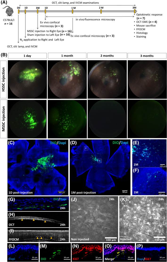
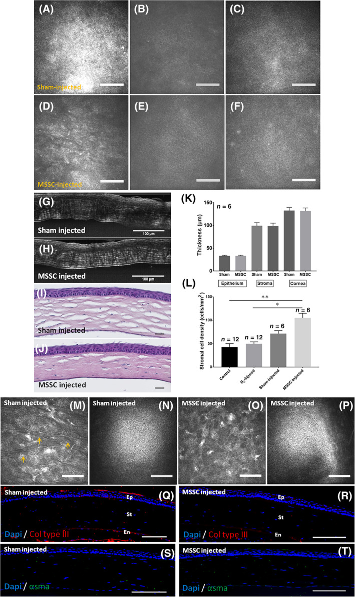
Corneal scarring associated with various corneal conditions is a leading cause of blindness worldwide. The present study aimed to test the hypothesis that corneal stromal stem cells have a therapeutic effect and are able to restore the extracellular matrix organization and corneal transparency in vivo. We first developed a mouse model of corneal stromal scar induced by liquid nitrogen (N ) application. We then reversed stromal scarring by injecting mouse or human corneal stromal stem cells in injured cornea. To characterize the mouse model developed in this study and the therapeutic effect of corneal stromal stem cells, we used a combination of in vivo (slit lamp, optical coherence tomography, in vivo confocal microscopy, optical coherence tomography shear wave elastography, and optokinetic tracking response) and ex vivo (full field optical coherence microscopy, flow cytometry, transmission electron microscopy, and histology) techniques. The mouse model obtained features early inflammation, keratocyte apoptosis, keratocyte transformation into myofibroblasts, collagen type III synthesis, impaired stromal ultrastructure, corneal stromal haze formation, increased corneal rigidity, and impaired visual acuity. Injection of stromal stem cells in N -injured cornea resulted in improved corneal transparency associated with corneal stromal stem cell migration and growth in the recipient stroma, absence of inflammatory response, recipient corneal epithelial cell growth, decreased collagen type III stromal content, restored stromal ultrastructure, decreased stromal haze, decreased corneal rigidity, and improved vision. Our study demonstrates the ability of corneal stromal stem cells to promote regeneration of transparent stromal tissue after corneal scarring induced by liquid nitrogen.
角膜瘢痕与各种角膜疾病有关,是全球致盲的主要原因。本研究旨在检验角膜基质干细胞具有治疗作用并能够恢复体内细胞外基质组织和角膜透明度的假说。我们首先通过液氮(N)应用开发了一种角膜基质瘢痕的小鼠模型。然后,我们通过向受损角膜注射小鼠或人角膜基质干细胞来逆转基质瘢痕。为了描述本研究中开发的小鼠模型和角膜基质干细胞的治疗效果,我们结合了体内(裂隙灯、光学相干断层扫描、活体共聚焦显微镜、光学相干断层扫描剪切波弹性成像和视觉追踪反应)和体外(全场光学相干显微镜、流式细胞术、透射电子显微镜和组织学)技术。获得的小鼠模型具有早期炎症、角膜细胞凋亡、角膜细胞向肌成纤维细胞转化、III 型胶原合成、基质超微结构受损、角膜基质混浊形成、角膜硬度增加和视力受损等特征。将基质干细胞注射到 N 损伤的角膜中,可改善角膜透明度,与受体基质中基质干细胞的迁移和生长有关,无炎症反应,受体角膜上皮细胞生长,III 型胶原基质含量减少,基质超微结构恢复,基质混浊减少,角膜硬度降低,视力提高。我们的研究表明,角膜基质干细胞具有在液氮诱导的角膜瘢痕后促进透明基质组织再生的能力。