Division of Cardiology, Department of Pediatrics, Medical College of Wisconsin, Children's Hospital of Wisconsin, Herma Heart Institute, Milwaukee, Wisconsin.
Division of Neonatology, Department of Pediatrics, Medical College of Wisconsin, Children's Hospital of Wisconsin, Milwaukee, Wisconsin.
Semin Thorac Cardiovasc Surg. 2020;32(4):980-987. doi: 10.1053/j.semtcvs.2020.03.004. Epub 2020 May 7.
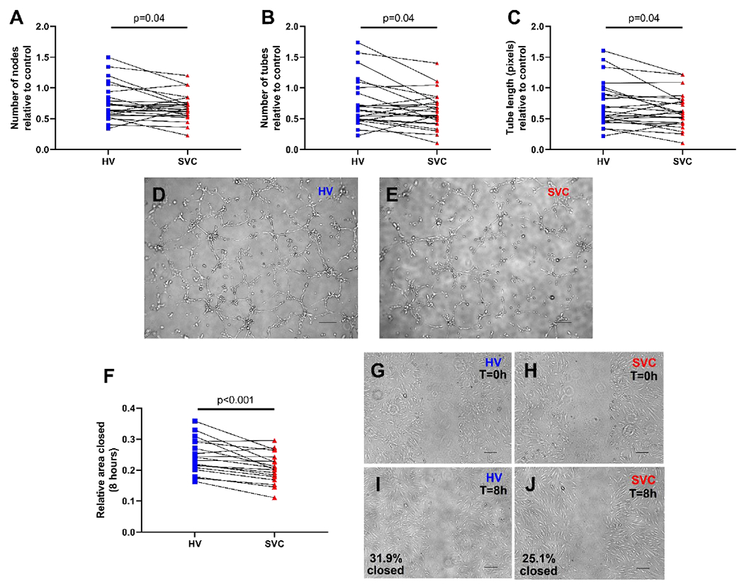
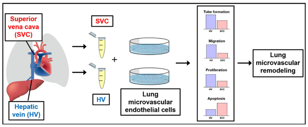
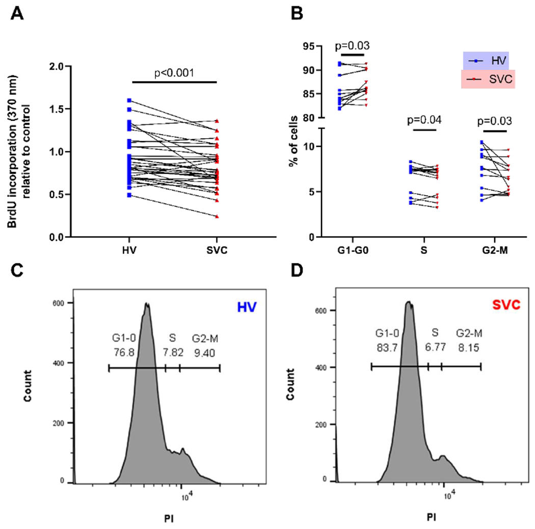
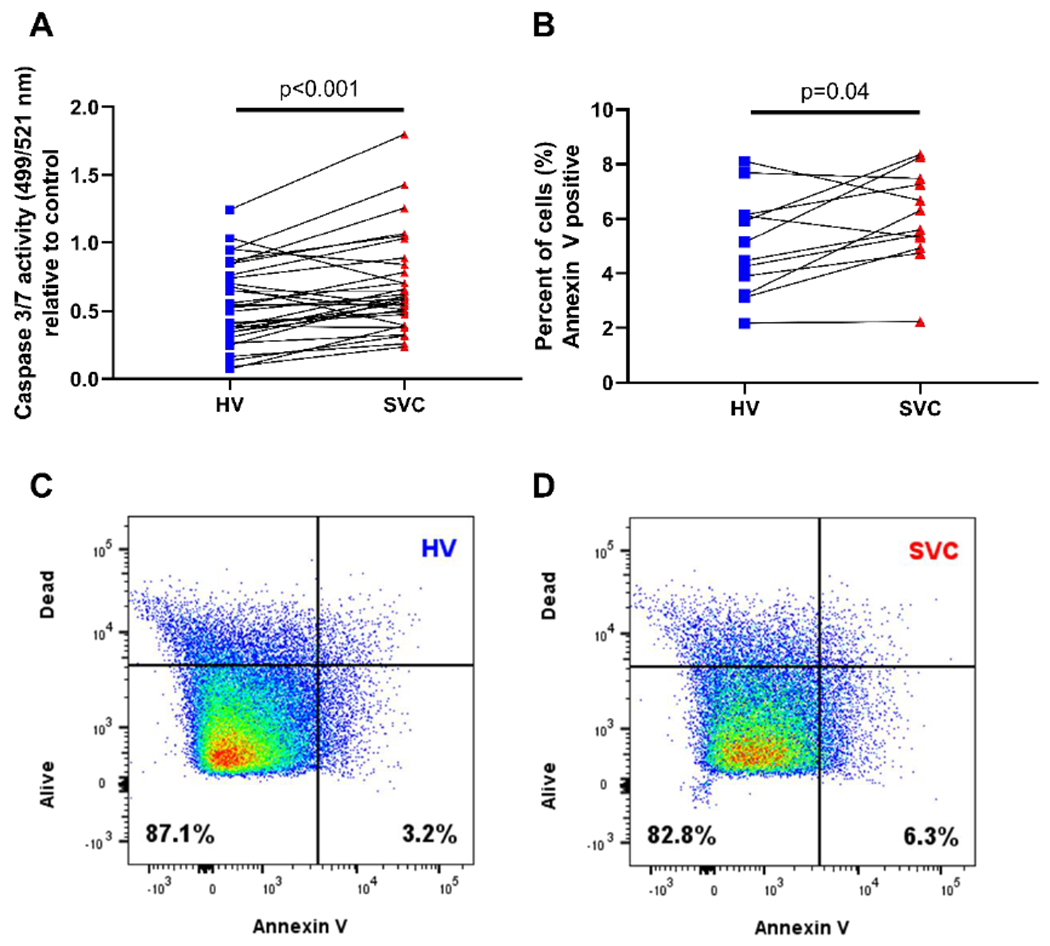
To improve our understanding of pulmonary arteriovenous malformations in univentricular congenital heart disease, our objective was to identify the effects of hepatic vein and superior vena cava constituents on lung microvascular endothelial cells independent of blood flow. Paired blood samples were collected from the hepatic vein and superior vena cava in children 0-10 years old undergoing cardiac catheterization. Isolated serum was subsequently used for in vitro endothelial cell assays. Angiogenic activity was assessed using tube formation and scratch migration. Endothelial cell survival was assessed using proliferation (BrdU incorporation, cell cycle analysis) and apoptosis (caspase 3/7 activity, Annexin-V labeling). Data were analyzed using Wilcoxon signed-rank test and repeated measures analysis. Upon incubating lung microvascular endothelial cells with 10% patient serum, hepatic vein serum increases angiogenic activity (tube formation, P = 0.04, n = 24; migration, P< 0.001, n = 18), increases proliferation (BrdU, P < 0.001, n = 32; S-phase, P = 0.04, n = 13), and decreases apoptosis (caspase 3/7, P < 0.001, n = 32; Annexin-V, P = 0.04, n = 12) compared to superior vena cava serum. Hepatic vein serum regulates lung microvascular endothelial cells by increasing angiogenesis and survival in vitro. Loss of hepatic vein serum signaling in the lung microvasculature may promote maladaptive lung microvascular remodeling and pulmonary arteriovenous malformations.
为了增进我们对单心室先天性心脏病中肺动静脉畸形的理解,我们的目的是在不考虑血流的情况下,确定肝静脉和上腔静脉成分对肺微血管内皮细胞的影响。对 0-10 岁行心导管术的儿童采集肝静脉和上腔静脉的配对血样。随后用分离的血清进行体外内皮细胞检测。通过管形成和划痕迁移评估血管生成活性。通过增殖(BrdU 掺入、细胞周期分析)和凋亡(caspase 3/7 活性、Annexin-V 标记)评估内皮细胞存活。使用 Wilcoxon 符号秩检验和重复测量分析来分析数据。在用 10%患者血清孵育肺微血管内皮细胞后,肝静脉血清增加血管生成活性(管形成,P=0.04,n=24;迁移,P<0.001,n=18),增加增殖(BrdU,P<0.001,n=32;S 期,P=0.04,n=13),并减少凋亡(caspase 3/7,P<0.001,n=32;Annexin-V,P=0.04,n=12)与上腔静脉血清相比。肝静脉血清通过增加体外血管生成和存活来调节肺微血管内皮细胞。肺微血管内皮细胞中肝静脉血清信号的丧失可能促进肺微血管重塑和肺动静脉畸形的适应性不良。