• 文献检索
  • 文档翻译
  • 深度研究
  • 学术资讯
  • Suppr Zotero 插件Zotero 插件
  • 邀请有礼
  • 套餐&价格
  • 历史记录
应用&插件
Suppr Zotero 插件Zotero 插件浏览器插件Mac 客户端Windows 客户端微信小程序
定价
高级版会员购买积分包购买API积分包
服务
文献检索文档翻译深度研究API 文档MCP 服务
关于我们
关于 Suppr公司介绍联系我们用户协议隐私条款
关注我们

Suppr 超能文献

核心技术专利:CN118964589B侵权必究
粤ICP备2023148730 号-1Suppr @ 2026

文献检索

告别复杂PubMed语法,用中文像聊天一样搜索,搜遍4000万医学文献。AI智能推荐,让科研检索更轻松。

立即免费搜索

文件翻译

保留排版,准确专业,支持PDF/Word/PPT等文件格式,支持 12+语言互译。

免费翻译文档

深度研究

AI帮你快速写综述,25分钟生成高质量综述,智能提取关键信息,辅助科研写作。

立即免费体验

共聚焦激光拉曼显微镜鉴定口腔龈下生物膜模型中的细菌。

Confocal Raman microscopy to identify bacteria in oral subgingival biofilm models.

机构信息

Fraunhofer Institute for Interfacial Engineering and Biotechnology, Stuttgart, Germany.

Procter & Gamble, Egham, England, United Kingdom.

出版信息

PLoS One. 2020 May 11;15(5):e0232912. doi: 10.1371/journal.pone.0232912. eCollection 2020.

DOI:10.1371/journal.pone.0232912
PMID:32392236
原文链接:https://pmc.ncbi.nlm.nih.gov/articles/PMC7213720/
Abstract

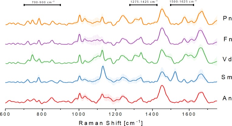
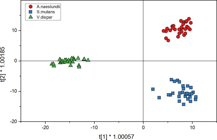
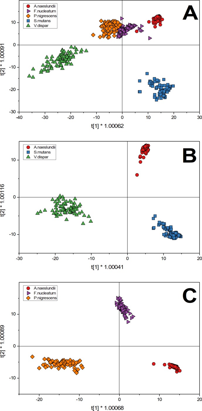
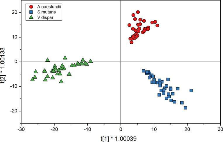
The study of oral disease progression, in relation to the accumulation of subgingival biofilm in gingivitis and periodontitis is limited, due to either the ability to monitor plaque in vitro. When compared, optical spectroscopic techniques offer advantages over traditional destructive or biofilm staining approaches, making it a suitable alternative for the analysis and continued development of three-dimensional structures. In this work, we have developed a confocal Raman spectroscopy analysis approach towards in vitro subgingival plaque models. The main objective of this study was to develop a method for differentiating multiple oral subgingival bacterial species in planktonic and biofilm conditions, using confocal Raman microscopy. Five common subgingival bacteria (Fusobacterium nucleatum, Streptococcus mutans, Veillonella dispar, Actinomyces naeslundii and Prevotella nigrescens) were used and differentiated using a 2-way orthogonal Partial Least Square with Discriminant Analysis (O2PLS-DA) for the collected spectral data. In addition to planktonic growth, mono-species biofilms cultured using the 'Zürich Model' were also analyzed. The developed method was successfully used to predict planktonic and mono-species biofilm species in a cross validation setup. The results show differences in the presence and absence of chemical bands within the Raman spectra. The O2PLS-DA model was able to successfully predict 100% of all tested planktonic samples and 90% of all mono-species biofilm samples. Using this approach we have shown that Confocal Raman microscopy can analyse and predict the identity of planktonic and mono-species biofilm species, thus enabling its potential as a technique to map oral multi-species biofilm models.

摘要

关于牙龈炎和牙周炎中龈下生物膜积累与口腔疾病进展的研究,由于体外监测牙菌斑的能力有限。与传统的破坏性或生物膜染色方法相比,光学光谱技术具有优势,使其成为分析和继续开发三维结构的合适替代方法。在这项工作中,我们开发了一种用于体外龈下牙菌斑模型的共焦拉曼光谱分析方法。本研究的主要目的是开发一种方法,用于区分浮游和生物膜条件下的多种口腔龈下细菌,使用共焦拉曼显微镜。使用了五种常见的龈下细菌(核梭杆菌、变形链球菌、差异韦荣球菌、奈瑟氏放线菌和变黑普雷沃菌),并使用收集的光谱数据的双向正交偏最小二乘判别分析(O2PLS-DA)进行区分。除了浮游生长外,还使用“苏黎世模型”培养单种生物膜。开发的方法成功地用于在交叉验证设置中预测浮游和单种生物膜物种。结果表明,拉曼光谱中存在和不存在化学带的差异。O2PLS-DA 模型能够成功预测所有测试浮游样本的 100%和所有单种生物膜样本的 90%。通过这种方法,我们已经表明共聚焦拉曼显微镜可以分析和预测浮游和单种生物膜物种的身份,从而使其有可能成为一种技术来绘制口腔多物种生物膜模型。

https://cdn.ncbi.nlm.nih.gov/pmc/blobs/604c/7213720/a17c63a07229/pone.0232912.g004.jpg
https://cdn.ncbi.nlm.nih.gov/pmc/blobs/604c/7213720/770dff7562af/pone.0232912.g001.jpg
https://cdn.ncbi.nlm.nih.gov/pmc/blobs/604c/7213720/5fc18d7cea46/pone.0232912.g002.jpg
https://cdn.ncbi.nlm.nih.gov/pmc/blobs/604c/7213720/9e6d01780a9e/pone.0232912.g003.jpg
https://cdn.ncbi.nlm.nih.gov/pmc/blobs/604c/7213720/a17c63a07229/pone.0232912.g004.jpg

相似文献

1
Confocal Raman microscopy to identify bacteria in oral subgingival biofilm models.共聚焦激光拉曼显微镜鉴定口腔龈下生物膜模型中的细菌。
PLoS One. 2020 May 11;15(5):e0232912. doi: 10.1371/journal.pone.0232912. eCollection 2020.
2
Structure, viability and bacterial kinetics of an in vitro biofilm model using six bacteria from the subgingival microbiota.使用龈下微生物群中的六种细菌构建体外生物膜模型的结构、活力和细菌动力学。
J Periodontal Res. 2011 Apr;46(2):252-60. doi: 10.1111/j.1600-0765.2010.01341.x. Epub 2011 Jan 25.
3
Validation of a quantitative real-time PCR assay and comparison with fluorescence microscopy and selective agar plate counting for species-specific quantification of an in vitro subgingival biofilm model.定量实时 PCR 检测方法的验证及其与荧光显微镜和选择性琼脂平板计数方法比较,用于体外龈下生物膜模型的种特异性定量。
J Periodontal Res. 2013 Aug;48(4):517-26. doi: 10.1111/jre.12034. Epub 2012 Dec 30.
4
Colonisation of gingival epithelia by subgingival biofilms in vitro: role of "red complex" bacteria.龈下生物膜在体外对牙龈上皮的定植:“红色复合体”细菌的作用
Arch Oral Biol. 2014 Sep;59(9):977-86. doi: 10.1016/j.archoralbio.2014.05.023. Epub 2014 Jun 4.
5
Mapping of a Subgingival Dual-Species Biofilm Model Using Confocal Raman Microscopy.使用共聚焦拉曼显微镜对龈下双物种生物膜模型进行映射分析。
Front Microbiol. 2021 Oct 5;12:729720. doi: 10.3389/fmicb.2021.729720. eCollection 2021.
6
Characterization and application of a flow system for in vitro multispecies oral biofilm formation.用于体外多物种口腔生物膜形成的流动系统的表征与应用
J Periodontal Res. 2014 Jun;49(3):323-32. doi: 10.1111/jre.12110. Epub 2013 Jul 1.
7
Species and Subspecies Differentially Affect the Composition and Architecture of Supra- and Subgingival Biofilms Models.物种和亚种对龈上和龈下生物膜模型的组成和结构有不同影响。
Front Microbiol. 2019 Jul 30;10:1716. doi: 10.3389/fmicb.2019.01716. eCollection 2019.
8
Impact of early colonizers on in vitro subgingival biofilm formation.早期定植菌对体外龈下生物膜形成的影响。
PLoS One. 2013 Dec 5;8(12):e83090. doi: 10.1371/journal.pone.0083090. eCollection 2013.
9
Quantitation of biofilm and planktonic life forms of coexisting periodontal species.共生牙周物种生物膜和浮游生物形式的定量分析。
Anaerobe. 2015 Oct;35(Pt A):13-20. doi: 10.1016/j.anaerobe.2015.04.013. Epub 2015 Apr 26.
10
The in vivo dynamics of Streptococcus spp., Actinomyces naeslundii, Fusobacterium nucleatum and Veillonella spp. in dental plaque biofilm as analysed by five-colour multiplex fluorescence in situ hybridization.通过五色多重荧光原位杂交分析牙菌斑生物膜中链球菌属、内氏放线菌、具核梭杆菌和韦荣氏菌属的体内动态。
J Med Microbiol. 2007 May;56(Pt 5):681-687. doi: 10.1099/jmm.0.47094-0.

引用本文的文献

1
The role of salivary metabolomics in chronic periodontitis: bridging oral and systemic diseases.唾液代谢组学在慢性牙周炎中的作用:连接口腔与全身疾病
Metabolomics. 2025 Feb 7;21(1):24. doi: 10.1007/s11306-024-02220-0.
2
Characterization and morphological methods for oral biofilm visualization: where are we nowadays?口腔生物膜可视化的表征与形态学方法:我们如今处于什么阶段?
AIMS Microbiol. 2024 Jun 6;10(2):391-414. doi: 10.3934/microbiol.2024020. eCollection 2024.
3
Rapid discrimination of four Salmonella enterica serovars: A performance comparison between benchtop and handheld Raman spectrometers.

本文引用的文献

1
Biofilm formation of clinically important microorganisms on 2D and 3D poly (methyl methacrylate) substrates: A surface-enhanced Raman scattering study.临床重要微生物在 2D 和 3D 聚甲基丙烯酸甲酯基底上的生物膜形成:表面增强拉曼散射研究。
Colloids Surf B Biointerfaces. 2020 Apr;188:110765. doi: 10.1016/j.colsurfb.2019.110765. Epub 2020 Jan 13.
2
Species and Subspecies Differentially Affect the Composition and Architecture of Supra- and Subgingival Biofilms Models.物种和亚种对龈上和龈下生物膜模型的组成和结构有不同影响。
Front Microbiol. 2019 Jul 30;10:1716. doi: 10.3389/fmicb.2019.01716. eCollection 2019.
3
四种肠炎沙门氏菌血清型的快速鉴别:台式与手持式拉曼光谱仪的性能比较
J Cell Mol Med. 2024 Apr;28(8):e18292. doi: 10.1111/jcmm.18292.
4
Omics for deciphering oral microecology.组学解析口腔微生态。
Int J Oral Sci. 2024 Jan 9;16(1):2. doi: 10.1038/s41368-023-00264-x.
5
Open-sourced Raman spectroscopy data processing package implementing a baseline removal algorithm validated from multiple datasets acquired in human tissue and biofluids.开源拉曼光谱数据分析处理软件包,实现了一种基线去除算法,该算法在人体组织和生物流体的多个数据集上进行了验证。
J Biomed Opt. 2023 Feb;28(2):025002. doi: 10.1117/1.JBO.28.2.025002. Epub 2023 Feb 21.
6
Identification of foodborne pathogenic bacteria using confocal Raman microspectroscopy and chemometrics.利用共聚焦拉曼光谱和化学计量学鉴定食源性病原体细菌
Front Microbiol. 2022 Nov 7;13:874658. doi: 10.3389/fmicb.2022.874658. eCollection 2022.
7
Recent progress in analyzing the spatial structure of the human microbiome: distinguishing biogeography and architecture in the oral and gut communities.人类微生物组空间结构分析的最新进展:区分口腔和肠道群落中的生物地理学与结构
Curr Opin Endocr Metab Res. 2021 Jun;18:275-283. doi: 10.1016/j.coemr.2021.04.005. Epub 2021 Apr 26.
8
Raman Spectroscopy: A Potential Diagnostic Tool for Oral Diseases.拉曼光谱学:口腔疾病的潜在诊断工具。
Front Cell Infect Microbiol. 2022 Feb 4;12:775236. doi: 10.3389/fcimb.2022.775236. eCollection 2022.
9
Mapping of a Subgingival Dual-Species Biofilm Model Using Confocal Raman Microscopy.使用共聚焦拉曼显微镜对龈下双物种生物膜模型进行映射分析。
Front Microbiol. 2021 Oct 5;12:729720. doi: 10.3389/fmicb.2021.729720. eCollection 2021.
10
Fast label-free identification of bacteria by synchronous fluorescence of amino acids.基于氨基酸同步荧光的快速无标记细菌鉴定
Anal Bioanal Chem. 2021 Nov;413(27):6857-6866. doi: 10.1007/s00216-021-03642-8. Epub 2021 Sep 7.
Dissecting the Metabolic Phenotype of the Antihypertensive Effects of Five Species on Spontaneously Hypertensive Rats.
剖析五种药物对自发性高血压大鼠降压作用的代谢表型
Front Pharmacol. 2019 Jul 30;10:845. doi: 10.3389/fphar.2019.00845. eCollection 2019.
4
The Recent Advances in Raman Microscopy and Imaging Techniques for Biosensors.用于生物传感器的拉曼显微镜和成像技术的最新进展。
Biosensors (Basel). 2019 Feb 12;9(1):25. doi: 10.3390/bios9010025.
5
Soil Particles and Phenanthrene Interact in Defining the Metabolic Profile of G7: A Vibrational Spectroscopy Approach.土壤颗粒与菲在定义G7的代谢谱中的相互作用:一种振动光谱方法
Front Microbiol. 2018 Dec 4;9:2999. doi: 10.3389/fmicb.2018.02999. eCollection 2018.
6
Fast discrimination of bacteria using a filter paper-based SERS platform and PLS-DA with uncertainty estimation.基于滤纸的 SERS 平台和具有不确定性估计的 PLS-DA 快速鉴别细菌。
Anal Bioanal Chem. 2019 Jan;411(3):705-713. doi: 10.1007/s00216-018-1485-9. Epub 2018 Nov 19.
7
Raman spectroscopic analysis of the molecular composition of oral cavity squamous cell carcinoma and healthy tongue tissue.口腔鳞状细胞癌和健康舌组织的分子组成的喇曼光谱分析。
Analyst. 2018 Aug 20;143(17):4090-4102. doi: 10.1039/c7an02106b.
8
Rapid identification of staphylococci by Raman spectroscopy.利用拉曼光谱快速鉴定葡萄球菌。
Sci Rep. 2017 Nov 1;7(1):14846. doi: 10.1038/s41598-017-13940-w.
9
Characterization and Discrimination of Gram-Positive Bacteria Using Raman Spectroscopy with the Aid of Principal Component Analysis.利用拉曼光谱并借助主成分分析对革兰氏阳性菌进行表征与鉴别
Nanomaterials (Basel). 2017 Sep 1;7(9):248. doi: 10.3390/nano7090248.
10
Raman spectroscopy uncovers biochemical tissue-related features of extracellular vesicles from mesenchymal stromal cells.拉曼光谱揭示了间充质基质细胞来源的细胞外囊泡的与生化组织相关的特征。
Sci Rep. 2017 Aug 29;7(1):9820. doi: 10.1038/s41598-017-10448-1.