Department of Normal and Clinical Anatomy, Anatomy and Histology, Medical University of Lodz, Poland.
Department of Neurosurgery, Tulane Center for Clinical Neurosciences, Tulane University School of Medicine, New Orleans, LA, USA.
Biomed Res Int. 2020 Apr 30;2020:9037693. doi: 10.1155/2020/9037693. eCollection 2020.
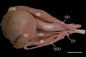
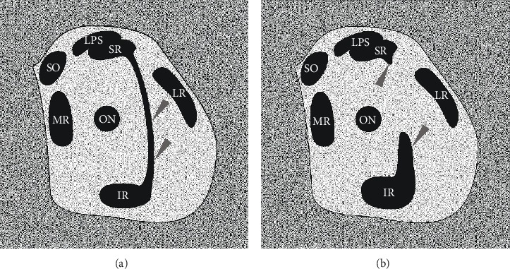
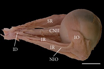
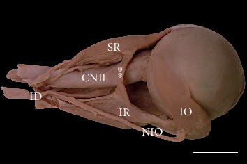
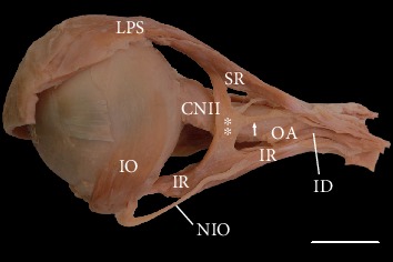
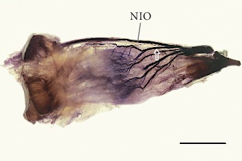
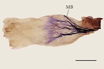
A comparison of the superior and inferior rectus muscles was performed to determine whether they have similar structures and innervation attributable to their participation in the same type of, although antagonistic, eye movements. The study was conducted on 70 cadaveric hemiheads, and the anatomical variations in the superior and inferior rectus muscles were assessed. Sihler's whole mount nerve staining technique was used on 20 isolated superior and 20 isolated inferior rectus muscle specimens to visualize the intramuscular distribution of the oculomotor nerve subbranches. In two cases (~2.8%), variant muscular slips were found that connected the superior and inferior rectus muscles. In 80% of cases, muscular branches arising directly from the inferior branch of the oculomotor nerve innervated the inferior rectus muscle, while in 20% of cases, the nerve to the inferior oblique muscle pierced the inferior rectus muscle and provided its innervation. In 15 of 70 specimens (21.4%), a branch to the levator palpebrae superioris muscle pierced the superior rectus muscle. The distance between the specific rectus muscle's insertion and the anterior-most terminations of the nerves' subbranches with reference to the muscle's total length ranged from 26.9% to 47.2% for the inferior rectus and from 34.8% to 46.6% for the superior rectus, respectively. The superior rectus muscle is slightly longer and its insertion is farther from the limbus of the cornea than is the inferior rectus muscle. Both muscles share a common general pattern of intramuscular nerve subbranches' arborization, with characteristic Y-shaped ramifications that form the terminal nerve plexus located near half of the muscles' length. Unexpected anatomical variations of the extraocular muscles may be relevant during orbital imaging or surgical procedures.
对眼直肌进行了比较,以确定它们是否具有相似的结构和神经支配,这归因于它们参与了相同类型的,尽管是拮抗的,眼球运动。本研究在 70 个头半标本上进行,评估了上直肌和下直肌的解剖变异。使用 Sihler 全肌肉神经染色技术对 20 个分离的上直肌和 20 个分离的下直肌标本进行了研究,以可视化动眼神经分支在肌肉内的分布。在两种情况下(约 2.8%),发现了连接上直肌和下直肌的变异肌束。在 80%的情况下,直接发自动眼神经下支的肌支支配下直肌,而在 20%的情况下,支配下斜肌的神经穿过下直肌并为其提供神经支配。在 70 个标本中的 15 个(21.4%),提上睑肌的分支穿过上直肌。特定直肌止点与神经分支最前端的距离与肌肉总长度的关系,下直肌为 26.9%至 47.2%,上直肌为 34.8%至 46.6%。上直肌略长,其止点距角膜缘较下直肌远。两块肌肉都有共同的肌内神经分支分支模式,具有特征性的 Y 形分支,形成位于肌肉长度一半附近的终末神经丛。眼外肌的意外解剖变异可能在眼眶成像或手术过程中具有重要意义。