• 文献检索
  • 文档翻译
  • 深度研究
  • 学术资讯
  • Suppr Zotero 插件Zotero 插件
  • 邀请有礼
  • 套餐&价格
  • 历史记录
应用&插件
Suppr Zotero 插件Zotero 插件浏览器插件Mac 客户端Windows 客户端微信小程序
定价
高级版会员购买积分包购买API积分包
服务
文献检索文档翻译深度研究API 文档MCP 服务
关于我们
关于 Suppr公司介绍联系我们用户协议隐私条款
关注我们

Suppr 超能文献

核心技术专利:CN118964589B侵权必究
粤ICP备2023148730 号-1Suppr @ 2026

文献检索

告别复杂PubMed语法,用中文像聊天一样搜索,搜遍4000万医学文献。AI智能推荐,让科研检索更轻松。

立即免费搜索

文件翻译

保留排版,准确专业,支持PDF/Word/PPT等文件格式,支持 12+语言互译。

免费翻译文档

深度研究

AI帮你快速写综述,25分钟生成高质量综述,智能提取关键信息,辅助科研写作。

立即免费体验

化疗放疗后早期弥散加权磁共振成像对头颈部鳞状细胞癌复发或肿瘤进展的预测价值

Predictive Value of Early Post-Treatment Diffusion-Weighted MRI for Recurrence or Tumor Progression of Head and Neck Squamous Cell Carcinoma Treated with Chemo-Radiotherapy.

作者信息

Brenet Esteban, Barbe Coralie, Hoeffel Christine, Dubernard Xavier, Merol Jean-Claude, Fath Léa, Servagi-Vernat Stéphanie, Labrousse Marc

机构信息

Department of Oto-Rhino-Laryngology, Head and Neck Surgery, Robert Debré University Hospital, 51100 Reims, France.

Clinical Research Unit, Robert Debré University Hospital, 51100 Reims, France.

出版信息

Cancers (Basel). 2020 May 14;12(5):1234. doi: 10.3390/cancers12051234.

DOI:10.3390/cancers12051234
PMID:32422975
原文链接:https://pmc.ncbi.nlm.nih.gov/articles/PMC7281260/
Abstract

AIMS

To investigate the predictive capacity of early post-treatment diffusion-weighted magnetic resonance imaging (MRI) for recurrence or tumor progression in patients with no tumor residue after chemo-radiotherapy (CRT) for head and neck squamous cell carcinoma, and, to assess the predictive capacity of pre-treatment diffusion-weighted MRI for persistent tumor residue post-CRT.

MATERIALS AND METHOD

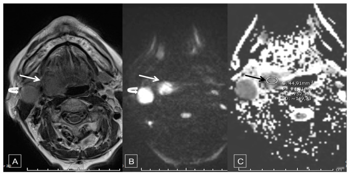
A single center cohort study was performed in one French hospital. All patients with squamous cell carcinoma receiving CRT (no surgical indication) were included. Two diffusion-weighted MRI were performed: one within 8 days before CRT and one 3 months after completing CRT with determination of median tumor apparent diffusion coefficient (ADC).

MAIN OUTCOME

The primary endpoint was progression-free survival.

RESULTS

59 patients were included prior to CRT and 46 (78.0%) completed CRT. A post-CRT tumor residue was found in 19/46 (41.3%) patients. In univariate analysis, initial ADC was significantly lower in patients with residue post CRT (0.56 ± 0.11 versus 0.79 ± 0.13; < 0.001). When initial ADC was dichotomized at the median, initial ADC lower than 0.7 was significantly more frequent in patients with residue post CRT (73.7% versus 11.1%, < 0.0001). In multivariate analysis, only initial ADC lower than 0.7 was significantly associated with tumor residue (OR = 22.6; IC [4.9-103.6], < 0.0001). Among 26 patients without tumor residue after CRT and followed up until 12 months, 6 (23.1%) presented recurrence or progression. Only univariate analysis was performed due to a small number of events. The only factor significantly associated with disease progression or early recurrence was the delta ADC ( = 0.0009). When ADC variation was dichotomized at the median, patients with ADC variation greater than 0.7 had time of disease-free survival significantly longer than patients with ADC variation lower than 0.7 (377.5 [286-402] days versus 253 [198-370], < 0.0001). Conclusion and relevance: Diffusion-weighted MRI could be a technique that enables differentiation of patients with high potential for early recurrence for whom intensive post-CRT monitoring is mandatory. Prospective studies with more inclusions would be necessary to validate our results.

摘要

目的

探讨头颈部鳞状细胞癌放化疗(CRT)后无肿瘤残留患者早期治疗后扩散加权磁共振成像(MRI)对复发或肿瘤进展的预测能力,并评估治疗前扩散加权MRI对CRT后持续性肿瘤残留的预测能力。

材料与方法

在一家法国医院进行了一项单中心队列研究。纳入所有接受CRT(无手术指征)的鳞状细胞癌患者。进行了两次扩散加权MRI检查:一次在CRT前8天内,另一次在完成CRT后3个月,测定肿瘤表观扩散系数(ADC)中位数。

主要结局

主要终点是无进展生存期。

结果

59例患者在CRT前纳入,46例(78.0%)完成了CRT。19/46例(41.3%)患者发现CRT后有肿瘤残留。单因素分析显示,CRT后有残留的患者初始ADC显著更低(0.56±0.11对0.79±0.13;<0.001)。当初始ADC以中位数进行二分法划分时,CRT后有残留的患者初始ADC低于0.7的情况显著更常见(73.7%对11.1%,<0.0001)。多因素分析显示,只有初始ADC低于0.7与肿瘤残留显著相关(OR=22.6;IC[4.9 - 103.6],<0.0001)。在26例CRT后无肿瘤残留且随访至12个月的患者中,6例(23.1%)出现复发或进展。由于事件数量少,仅进行了单因素分析。与疾病进展或早期复发显著相关的唯一因素是ADC变化量(=0.0009)。当ADC变化量以中位数进行二分法划分时,ADC变化量大于0.7的患者无病生存期显著长于ADC变化量低于0.7的患者(377.5[286 - 402]天对253[198 - 370]天,<0.0001)。结论与相关性:扩散加权MRI可能是一种能够区分早期复发高风险患者的技术,对于这些患者,CRT后强化监测是必要的。需要更多纳入病例的前瞻性研究来验证我们的结果。

https://cdn.ncbi.nlm.nih.gov/pmc/blobs/1ebd/7281260/92f7d57763b4/cancers-12-01234-g004.jpg
https://cdn.ncbi.nlm.nih.gov/pmc/blobs/1ebd/7281260/71c79ba6f59f/cancers-12-01234-g001.jpg
https://cdn.ncbi.nlm.nih.gov/pmc/blobs/1ebd/7281260/7f6db94fd602/cancers-12-01234-g002.jpg
https://cdn.ncbi.nlm.nih.gov/pmc/blobs/1ebd/7281260/09ea038ae0b0/cancers-12-01234-g003.jpg
https://cdn.ncbi.nlm.nih.gov/pmc/blobs/1ebd/7281260/92f7d57763b4/cancers-12-01234-g004.jpg

相似文献

1
Predictive Value of Early Post-Treatment Diffusion-Weighted MRI for Recurrence or Tumor Progression of Head and Neck Squamous Cell Carcinoma Treated with Chemo-Radiotherapy.化疗放疗后早期弥散加权磁共振成像对头颈部鳞状细胞癌复发或肿瘤进展的预测价值
Cancers (Basel). 2020 May 14;12(5):1234. doi: 10.3390/cancers12051234.
2
Pretreatment ADC is not a prognostic factor for local recurrences in head and neck squamous cell carcinoma when clinical T-stage is known.当已知临床 T 分期时,预处理 ADC 不是头颈部鳞状细胞癌局部复发的预后因素。
Eur Radiol. 2020 Feb;30(2):1228-1231. doi: 10.1007/s00330-019-06426-y. Epub 2019 Sep 16.
3
Diffusion-weighted magnetic resonance imaging early after chemoradiotherapy to monitor treatment response in head-and-neck squamous cell carcinoma.放化疗后早期磁共振弥散加权成像监测头颈部鳞癌治疗反应
Int J Radiat Oncol Biol Phys. 2012 Mar 1;82(3):1098-107. doi: 10.1016/j.ijrobp.2011.02.044. Epub 2011 Apr 20.
4
Oesophageal squamous cell carcinoma: histogram-derived ADC parameters are not predictive of tumour response to chemoradiotherapy.食管鳞状细胞癌:直方图衍生 ADC 参数不能预测肿瘤对放化疗的反应。
Eur Radiol. 2018 Oct;28(10):4296-4305. doi: 10.1007/s00330-018-5439-6. Epub 2018 May 3.
5
Locally advanced rectal cancer: Qualitative and quantitative evaluation of diffusion-weighted MR imaging in the response assessment after neoadjuvant chemo-radiotherapy.局部进展期直肠癌:新辅助放化疗后反应评估中扩散加权磁共振成像的定性和定量评估
Eur J Radiol Open. 2016 Jul 18;3:145-52. doi: 10.1016/j.ejro.2016.06.003. eCollection 2016.
6
Predicting tumor recurrence in patients with cervical carcinoma treated with definitive chemoradiotherapy: value of quantitative histogram analysis on diffusion-weighted MR images.预测接受根治性放化疗的宫颈癌患者的肿瘤复发:扩散加权磁共振图像定量直方图分析的价值
Acta Radiol. 2017 Apr;58(4):481-488. doi: 10.1177/0284185116656492. Epub 2016 Jul 28.
7
Value of diffusion-weighted MRI and apparent diffusion coefficient measurements for predicting the response of locally advanced rectal cancer to neoadjuvant chemoradiotherapy.弥散加权 MRI 及其表观弥散系数测量在预测局部进展期直肠癌新辅助放化疗疗效中的价值。
Abdom Radiol (NY). 2016 Oct;41(10):1906-17. doi: 10.1007/s00261-016-0805-9.
8
A prospective, multi-centre trial of multi-parametric MRI as a biomarker in anal carcinoma.多参数 MRI 作为肛门癌生物标志物的前瞻性多中心研究。
Radiother Oncol. 2020 Mar;144:7-12. doi: 10.1016/j.radonc.2019.10.001. Epub 2019 Nov 8.
9
Investigation of volumetric apparent diffusion coefficient histogram analysis for assessing complete response and clinical outcomes following pre-operative chemoradiation treatment for rectal carcinoma.直肠癌术前放化疗后完全缓解及临床结局评估的容积表观扩散系数直方图分析研究。
Abdom Radiol (NY). 2017 May;42(5):1310-1318. doi: 10.1007/s00261-016-1010-6.
10
Integrating pretreatment diffusion weighted MRI into a multivariable prognostic model for head and neck squamous cell carcinoma.将治疗前扩散加权磁共振成像纳入头颈部鳞状细胞癌的多变量预后模型。
Radiother Oncol. 2014 Mar;110(3):429-34. doi: 10.1016/j.radonc.2014.01.004. Epub 2014 Mar 11.

引用本文的文献

1
PI3K/Akt1 Pathway Suppression by Quercetin-Doxorubicin Combination in Osteosarcoma Cell Line (MG-63 Cells).槲皮素-阿霉素联合用药对骨肉瘤细胞系(MG-63细胞)PI3K/Akt1信号通路的抑制作用
Medicina (Kaunas). 2025 Jul 25;61(8):1347. doi: 10.3390/medicina61081347.
2
The use of diffusion-weighted magnetic resonance imaging and parametric response mapping for disease outcome prediction in nasopharyngeal carcinoma.利用扩散加权磁共振成像和参数反应映射预测鼻咽癌的疾病转归
BMC Med Imaging. 2025 Jul 31;25(1):308. doi: 10.1186/s12880-025-01847-2.
3
Change in diffusion weighted imaging after induction chemotherapy outperforms RECIST guideline for long-term outcome prediction in advanced nasopharyngeal carcinoma.

本文引用的文献

1
Epidemiology of head and neck cancers: an update.头颈部癌症的流行病学:最新研究进展。
Curr Opin Oncol. 2020 May;32(3):178-186. doi: 10.1097/CCO.0000000000000629.
2
Can apparent diffusion coefficient (ADC) distinguish breast cancer from benign breast findings? A meta-analysis based on 13 847 lesions.表观扩散系数(ADC)能否区分乳腺癌与良性乳腺病变?基于 13847 个病灶的荟萃分析。
BMC Cancer. 2019 Oct 15;19(1):955. doi: 10.1186/s12885-019-6201-4.
3
Prognostic role of diffusion weighted and dynamic contrast-enhanced MRI in loco-regionally advanced head and neck cancer treated with concomitant chemoradiotherapy.
诱导化疗后弥散加权成像的变化在预测晚期鼻咽癌长期预后方面优于RECIST指南。
Cancer Imaging. 2025 Mar 12;25(1):32. doi: 10.1186/s40644-025-00854-4.
4
Can initial apparent diffusion coefficient and tumour volume predict future metastases in treatment-naive patients with laryngeal squamous cell carcinoma.初始表观扩散系数和肿瘤体积能否预测初治喉鳞状细胞癌患者未来的转移情况?
Pol J Radiol. 2024 May 24;89:e267-e272. doi: 10.5114/pjr/187675. eCollection 2024.
5
Applying multisequence MRI radiomics of the primary tumor and lymph node to predict HPV-related p16 status in patients with oropharyngeal squamous cell carcinoma.应用原发性肿瘤和淋巴结的多序列MRI影像组学预测口咽鳞状细胞癌患者的HPV相关p16状态。
Quant Imaging Med Surg. 2023 Apr 1;13(4):2234-2247. doi: 10.21037/qims-22-819. Epub 2023 Feb 9.
6
Imaging of human papilloma virus associated oropharyngeal squamous cell carcinoma and its impact on diagnosis, prognostication, and response assessment.人乳头瘤病毒相关口咽鳞状细胞癌的影像学表现及其对诊断、预后和反应评估的影响。
Br J Radiol. 2022 Sep 1;95(1138):20220149. doi: 10.1259/bjr.20220149. Epub 2022 Jun 28.
7
The ability of post-chemoradiotherapy DWI ADC and F-FDG SUV to predict treatment outcomes in head and neck cancer: impact of human papilloma virus oropharyngeal cancer status.放化疗后 DWI ADC 值和 F-FDG SUV 值预测头颈部鳞癌治疗效果的能力:人乳头瘤病毒阳性口咽癌状态的影响。
J Cancer Res Clin Oncol. 2021 Aug;147(8):2323-2336. doi: 10.1007/s00432-021-03662-y. Epub 2021 Jun 22.
弥散加权和动态对比增强 MRI 在同期放化疗治疗局部区域进展期头颈部癌症中的预后作用。
Radiol Oncol. 2019 Mar 3;53(1):39-48. doi: 10.2478/raon-2019-0010.
4
ADC-histogram analysis in head and neck squamous cell carcinoma. Associations with different histopathological features including expression of EGFR, VEGF, HIF-1α, Her 2 and p53. A preliminary study.头颈部鳞状细胞癌的ADC直方图分析。与不同组织病理学特征的关联,包括表皮生长因子受体(EGFR)、血管内皮生长因子(VEGF)、缺氧诱导因子-1α(HIF-1α)、人表皮生长因子受体2(Her 2)和p53的表达。一项初步研究。
Magn Reson Imaging. 2018 Dec;54:214-217. doi: 10.1016/j.mri.2018.07.013. Epub 2018 Sep 4.
5
Can diffusion-weighted imaging predict tumor grade and expression of Ki-67 in breast cancer? A multicenter analysis.弥散加权成像能否预测乳腺癌的肿瘤分级和 Ki-67 表达?一项多中心分析。
Breast Cancer Res. 2018 Jun 19;20(1):58. doi: 10.1186/s13058-018-0991-1.
6
Detection of loco-regional recurrence in malignant head and neck tumors: a comparison of CT, MRI, and FDG PET-CT.恶性头颈部肿瘤局部区域复发的检测:CT、MRI和FDG PET-CT的比较
Acta Radiol. 2019 Feb;60(2):186-195. doi: 10.1177/0284185118776504. Epub 2018 May 12.
7
Can Imaging Parameters Provide Information Regarding Histopathology in Head and Neck Squamous Cell Carcinoma? A Meta-Analysis.影像学参数能否提供有关头颈部鳞状细胞癌组织病理学的信息?一项荟萃分析。
Transl Oncol. 2018 Apr;11(2):498-503. doi: 10.1016/j.tranon.2018.02.004. Epub 2018 Mar 3.
8
Associations between apparent diffusion coefficient (ADC) and KI 67 in different tumors: a meta-analysis. Part 2: ADC.不同肿瘤中表观扩散系数(ADC)与Ki-67之间的关联:一项荟萃分析。第2部分:ADC
Oncotarget. 2018 Jan 4;9(9):8675-8680. doi: 10.18632/oncotarget.24006. eCollection 2018 Feb 2.
9
Associations between apparent diffusion coefficient (ADC) and KI 67 in different tumors: a meta-analysis. Part 1: ADC.不同肿瘤中表观扩散系数(ADC)与Ki-67之间的关联:一项荟萃分析。第1部分:ADC
Oncotarget. 2017 Aug 24;8(43):75434-75444. doi: 10.18632/oncotarget.20406. eCollection 2017 Sep 26.
10
Apparent Diffusion Coefficient Histograms of Human Papillomavirus-Positive and Human Papillomavirus-Negative Head and Neck Squamous Cell Carcinoma: Assessment of Tumor Heterogeneity and Comparison with Histopathology.人乳头瘤病毒阳性和人乳头瘤病毒阴性头颈部鳞状细胞癌的表观扩散系数直方图:肿瘤异质性评估及与组织病理学的比较。
AJNR Am J Neuroradiol. 2017 Nov;38(11):2153-2160. doi: 10.3174/ajnr.A5370. Epub 2017 Sep 14.