The First Clinical Medical College of Nanchang University, Nanchang, Jiangxi, China (mainland).
The First Afliated Hospital of Nanchang University, Nanchang, Jiangxi, China (mainland).
Med Sci Monit. 2020 May 19;26:e921606. doi: 10.12659/MSM.921606.
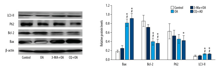
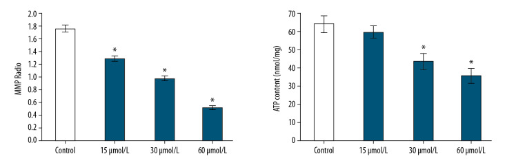
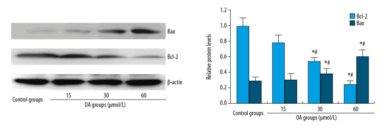
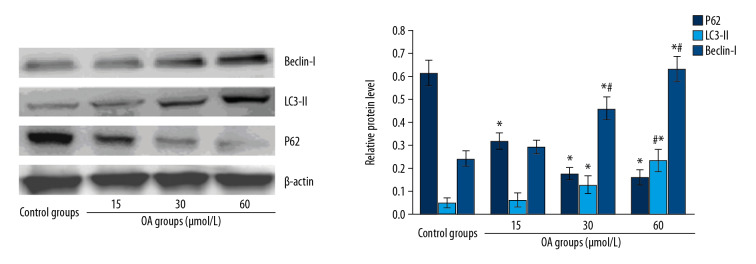
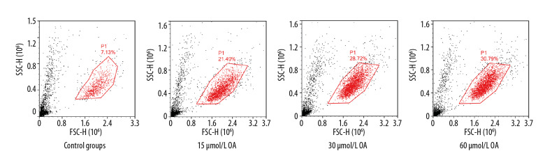
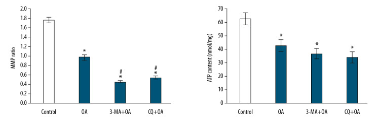
BACKGROUND Liver cancer is a common cancer with high morbidity and mortality. Due to the large toxic side effects of chemotherapeutic drugs and the overexpression of multidrug resistance genes in liver cancer, no effective chemotherapeutic drug has yet been found. Therefore, the search for a highly effective, low-toxic, and safe natural anticancer therapy is a hot issue. MATERIAL AND METHODS SMMC-7721 cells (a hepatocellular carcinoma cell line) were treated with different concentrations of oleanolic acid (OA) plus autophagy inhibitor 3-methyladenine (3-MA) (3-MA+OA) or chloroquine (CQ) plus OA (CQ+OA). We used MTT and Hoechst 33258 staining methods to determine the proliferation and apoptotic effect of OA on cells. Flow cytometry was used to detect apoptosis. Mitochondrial function was assessed by measuring mitochondrial membrane potential and adenosine triphosphate (ATP) concentration. To evaluate the ability of OA on apoptosis and autophagy mechanisms on SMMC 7721 cells, the related protein expression for apoptosis, autophagy, and the autophagic pathway were detected and analyzed by western blot. RESULTS OA can inhibit and induce apoptosis of SMMC-7721 in a dose-dependent manner. Compared with the control group, OA significantly reduced the intracellular mitochondrial membrane potential, and the intracellular ATP concentration was also significantly reduced. Moreover, OA reduced the expression of p-Akt and p-mTOR. The expression of p62 was decreased, and LC3-II and Beclin-1 protein expression levels increased. After inhibiting autophagy with 3-MA or CQ, compared with OA alone, cell mitochondrial membrane potential and ATP concentration were significantly reduced, cell p62 expression was reduced, and LC3-II expression was increased, apoptosis-related protein Bax protein was increased, and Bcl-2 protein was decreased, which suggested that 3-MA or CQ treatment increased OA-induced apoptosis of SMMC-7721 cells. This suggested that OA activated autophagy of SMMC-7721 cells in a protective autophagic manner. CONCLUSIONS The study findings suggest that OA combined with autophagy inhibitor 3-MA can better exert its anticancer effect.
肝癌是一种发病率和死亡率都很高的常见癌症。由于化疗药物的毒性副作用大,肝癌中多药耐药基因的过度表达,目前还没有发现有效的化疗药物。因此,寻找一种高效、低毒、安全的天然抗癌治疗方法是一个热点问题。
用不同浓度的齐墩果酸(OA)联合自噬抑制剂 3-甲基腺嘌呤(3-MA)(3-MA+OA)或氯喹(CQ)联合 OA(CQ+OA)处理 SMMC-7721 细胞(肝癌细胞系)。采用 MTT 和 Hoechst 33258 染色法测定 OA 对细胞增殖和凋亡的影响。流式细胞术检测细胞凋亡。通过测量线粒体膜电位和三磷酸腺苷(ATP)浓度来评估线粒体功能。为了评估 OA 对 SMMC 7721 细胞凋亡和自噬机制的作用能力,通过 Western blot 检测和分析凋亡、自噬和自噬途径的相关蛋白表达。
OA 可呈剂量依赖性抑制和诱导 SMMC-7721 细胞凋亡。与对照组相比,OA 显著降低了细胞内线粒体膜电位,细胞内 ATP 浓度也显著降低。此外,OA 降低了 p-Akt 和 p-mTOR 的表达。p62 的表达减少,LC3-II 和 Beclin-1 蛋白表达水平增加。用 3-MA 或 CQ 抑制自噬后,与 OA 单独作用相比,细胞线粒体膜电位和 ATP 浓度明显降低,细胞 p62 表达减少,LC3-II 表达增加,凋亡相关蛋白 Bax 蛋白增加,Bcl-2 蛋白减少,提示 3-MA 或 CQ 处理增加了 OA 诱导的 SMMC-7721 细胞凋亡。这表明 OA 以保护性自噬的方式激活了 SMMC-7721 细胞的自噬。
研究结果表明,OA 联合自噬抑制剂 3-MA 可以更好地发挥其抗癌作用。