Department of Prosthetic Dentistry, University Hospital, LMU Munich, Goethestraße 70, 80336, Munich, Germany.
Alpenpraxis Miesbach, Fraunhoferstr 10, 83714, Miesbach, Germany.
Clin Oral Investig. 2020 Dec;24(12):4511-4518. doi: 10.1007/s00784-020-03316-2. Epub 2020 May 20.
To evaluate the influence of intraoral scanning on the quality of preparations for all-ceramic single crowns.
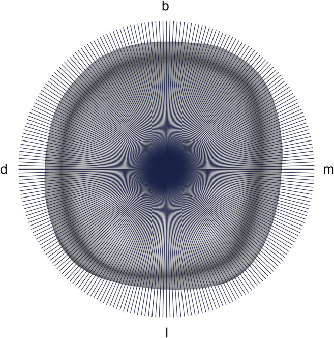
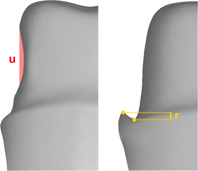
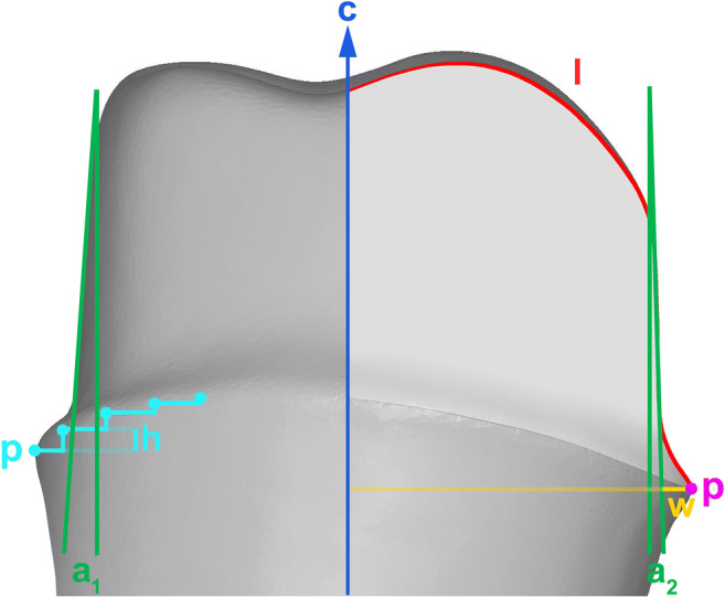
A total of 690 randomly selected and anonymized in vivo single crown preparations were examined. Three hundred twenty-three preparations were directly recorded with an intraoral scanner (group IS). Data from plaster casts digitized by a laboratory scanner (group ID; N = 367) served as control. Comparisons included convergence angle, marginal design, marginal substance reduction, homogeneity of the finish line, and undercuts. Evaluation was performed using fully automated specialized software. Data were analyzed applying Kolmogorov-Smirnov, Mann-Whitney U test, and Fisher's exact test. Level of significance was set at p < 0.05.
Convergence angle was above optimum in both groups, but significantly larger for group IS (p < 0.001). Marginal design was more ideal in group IS concerning the absence of featheredge design (p < 0.001) and reverse bevel (p = 0.211). Marginal substance reduction was closer to prerequisites for all-ceramic restorations in group IS (p < 0.001). Finish lines were more homogeneous in group IS regarding the uniformity of their course (p < 0.001). Undercuts were more frequently found in group ID than in group IS (p < 0.001).
Intraoral scanning of prepared teeth has positive impact on the quality of preparations for all-ceramic single crowns regarding marginal substance reduction, marginal design, homogeneity of the finish line, and undercuts.
Accurate preparation design represents a fundamental condition for success of ceramic crowns. Since there is potential for optimization, intraoral scanning might enhance preparation quality providing instant visual feedback.
评估口内扫描对全瓷单冠预备体质量的影响。
共检查了 690 个随机选择和匿名的体内单冠预备体。323 个预备体直接用口内扫描仪记录(IS 组)。由实验室扫描仪数字化的石膏模型数据(ID 组;N=367)作为对照。比较包括收敛角、边缘设计、边缘材料减少、边缘线均匀性和倒凹。使用全自动专用软件进行评估。使用 Kolmogorov-Smirnov、Mann-Whitney U 检验和 Fisher 精确检验进行数据分析。显著性水平设为 p<0.05。
两组的收敛角均高于最佳值,但 IS 组明显更大(p<0.001)。IS 组的边缘设计更理想,无羽状边缘设计(p<0.001)和反向斜面(p=0.211)。IS 组的边缘材料减少更接近全瓷修复的要求(p<0.001)。IS 组的边缘线更均匀,边缘线均匀性更好(p<0.001)。ID 组的倒凹比 IS 组更常见(p<0.001)。
口内扫描对全瓷单冠预备体的质量有积极影响,体现在边缘材料减少、边缘设计、边缘线均匀性和倒凹方面。
准确的预备设计是陶瓷冠成功的基本条件。由于存在优化的潜力,口内扫描可以通过提供即时的视觉反馈来提高预备质量。