Department of Radiology, Örebro university Hospital, Region Örebro län, S-70185 Örebro, Sweden.
Department of Radiology, Faculty of Medicine and Health, Örebro University, S-70182 Örebro, Sweden.
Br J Radiol. 2020 Jul;93(1111):20190995. doi: 10.1259/bjr.20190995. Epub 2020 May 21.
To perform CT pulmonary angiography (CTPA) using a minimal amount of iodinated contrast media.
47 patients (25 females) with mean age 69 years (range 41-82 years) referred for contrast-enhanced chest CT were prospectively included in this Phase IV clinical drug trial. All participants underwent a study specific CTPA in addition to the chest CT. The participants received 80 mg I/kg body weight Iohexol contrast media using a preparatory saline bolus, a dual flow contrast/saline bolus and a saline flush, and a scanner protocol with 80 kVp dual source high-pitch mode. Three readers independently assessed the image quality on the 3-point scale non-diagnostic, adequate or good-excellent image quality. Additionally, the pulmonary arterial contrast opacification was measured.
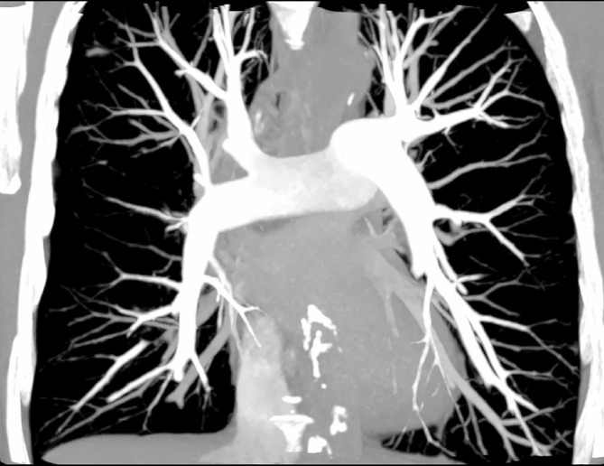
On average, the patients received 16.8 ml Iohexol 350 mg I/mL (range 12-20 ml). Mean patient weight was 71 kg (range 50-85 kg). Identically for all readers, pulmonary embolism (PE) was detected in 1/47 participants. The median number of examinations visually scored concerning pulmonary embolism as good-excellent was 47/47 (range 44-47); adequate 0/47 (0-3) and non-diagnostic 0/47 (range 0-0). The proportion adequate or better examinations was for all readers 47/47, 100% [95% confidence interval 92-100%]. The mean attenuation ± standard deviation in the pulmonary trunk was 325 ± 72 Hounsfield unit (range 165-531 Hounsfield unit).
Diagnostic CTPA with 17 ml contrast media is possible in non-obese patients using low kVp, high pitch and carefully designed contrast media administration.
By combining several procedures in a CTPA protocol, the contrast media dose can be minimized.
使用最小剂量的碘造影剂进行 CT 肺动脉造影(CTPA)。
本项 IV 期临床试验前瞻性纳入了 47 例(25 名女性)年龄 69 岁(41-82 岁)的患者,这些患者因怀疑患有胸部疾病而接受对比增强胸部 CT 检查。所有患者均接受特定的 CTPA 检查和胸部 CT 检查。患者使用 80mgI/kg 体重的碘海醇造影剂,采用预备盐水团注、双流速造影剂/盐水团注和盐水冲洗,以及 80kVp 双源高管电压高螺距模式的扫描仪方案。三位观察者独立对 3 分制(非诊断、充分、良好-优秀)的图像质量进行评估。此外,还对肺动脉对比度进行了测量。
平均每位患者接受 16.8ml 碘海醇 350mgI/ml(范围为 12-20ml)。患者平均体重为 71kg(范围为 50-85kg)。所有观察者均在 1/47 名患者中发现了肺栓塞(PE)。对所有观察者而言,47/47(范围 44-47)个肺动脉栓塞评估为良好-优秀,0/47(0-3)个为充分,0/47(0-0)个为非诊断。对于所有观察者,充分或更好的检查比例为 47/47,100%(95%置信区间 92-100%)。肺动脉干的平均衰减值±标准差为 325±72 亨氏单位(范围为 165-531 亨氏单位)。
在非肥胖患者中,通过结合 CTPA 方案中的多个步骤,使用低 kVp、高管电压和精心设计的造影剂给药方案,可实现 17ml 造影剂的诊断性 CTPA。
通过在 CTPA 方案中组合多个程序,可以最小化造影剂剂量。