Thome Janine, Densmore Maria, Koppe Georgia, Terpou Braeden, Théberge Jean, McKinnon Margaret C, Lanius Ruth A
Department of Psychiatry, Western University, London, Ontario, Canada.
Department of Theoretical Neuroscience, Central Institute of Mental Health Mannheim, Medical Faculty Mannheim, Heidelberg University, Mannheim, Germany.
Chronic Stress (Thousand Oaks). 2019 Sep 27;3:2470547019873663. doi: 10.1177/2470547019873663. eCollection 2019 Jan-Dec.
Brainstem and midbrain neuronal circuits that control innate, reflexive responses and arousal are increasingly recognized as central to the neurobiological framework of post-traumatic stress disorder (PTSD). The reticular activation system represents a fundamental neuronal circuit that plays a critical role not only in generating arousal but also in coordinating innate, reflexive responding. Accordingly, the present investigation aims to characterize the resting state functional connectivity of the reticular activation system in PTSD and its dissociative subtype.
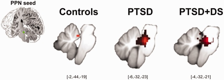
We investigated patterns of resting state functional connectivity of a central node of the reticular activation system, namely, the pedunculopontine nuclei, among individuals with PTSD (n = 77), its dissociative subtype (PTSD+DS; n = 48), and healthy controls (n = 51).
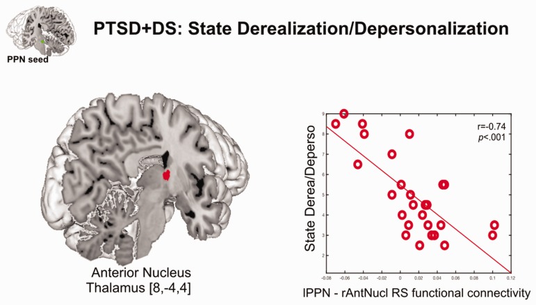
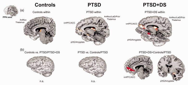
Participants with PTSD and PTSD+DS were characterized by within-group pedunculopontine nuclei resting state functional connectivity to brain regions involved in innate threat processing and arousal modulation (i.e., midbrain, amygdala, ventromedial prefrontal cortex). Critically, this pattern was most pronounced in individuals with PTSD+DS, as compared to both control and PTSD groups. As compared to participants with PTSD and controls, individuals with PTSD+DS showed enhanced pedunculopontine nuclei resting state functional connectivity to the amygdala and the parahippocampal gyrus as well as to the anterior cingulate and the ventromedial prefrontal cortex. No group differences emerged between PTSD and control groups. In individuals with PTSD+DS, state derealization/depersonalization was associated with reduced resting state functional connectivity between the left pedunculopontine nuclei and the anterior nucleus of the thalamus. Altered connectivity in these regions may restrict the thalamo-cortical transmission necessary to integrate internal and external signals at a cortical level and underlie, in part, experiences of depersonalization and derealization.
The present findings extend the current neurobiological model of PTSD and provide emerging evidence for the need to incorporate brainstem structures, including the reticular activation system, into current conceptualizations of PTSD and its dissociative subtype.
控制先天反射性反应和觉醒的脑干及中脑神经元回路,越来越被认为是创伤后应激障碍(PTSD)神经生物学框架的核心。网状激活系统是一个基本的神经元回路,不仅在产生觉醒方面起关键作用,还在协调先天反射性反应方面发挥重要作用。因此,本研究旨在描述PTSD及其分离亚型中网状激活系统的静息态功能连接特征。
我们研究了网状激活系统的一个中心节点,即脚桥核,在PTSD患者(n = 77)、其分离亚型(PTSD+DS;n = 48)和健康对照者(n = 51)中的静息态功能连接模式。
PTSD患者和PTSD+DS患者的特征是,组内脚桥核与参与先天威胁处理和觉醒调节的脑区(即中脑、杏仁核、腹内侧前额叶皮质)存在静息态功能连接。关键的是,与对照组和PTSD组相比,这种模式在PTSD+DS患者中最为明显。与PTSD患者和对照组相比,PTSD+DS患者的脚桥核与杏仁核、海马旁回以及前扣带回和腹内侧前额叶皮质的静息态功能连接增强。PTSD组和对照组之间未出现组间差异。在PTSD+DS患者中,现实解体/人格解体状态与左侧脚桥核和丘脑前核之间的静息态功能连接减少有关。这些区域连接性的改变可能会限制在皮质水平整合内部和外部信号所需的丘脑-皮质传递,并部分解释人格解体和现实解体的体验。
本研究结果扩展了当前的PTSD神经生物学模型,并为将包括网状激活系统在内的脑干结构纳入PTSD及其分离亚型的当前概念化提供了新的证据。