Clinic of Diagnostic and Interventional Radiology, Saarland University Medical Center, Homburg, Germany.
1st Department of Medicine (Cardiology, Angiology, Pulmonary and Intensive Care), University Medical Center Mannheim, Medical Faculty Mannheim, Heidelberg University, Mannheim, Germany.
PLoS One. 2020 May 29;15(5):e0233622. doi: 10.1371/journal.pone.0233622. eCollection 2020.
Quantified computed tomography (qCT) is known for correlations with airflow obstruction and fibrotic changes of the lung. However, as qCT studies often focus on diseased and elderly subjects, current literature lacks physiological qCT values during body development. We evaluated chest CT examinations of a healthy cohort, reaching from infancy to adulthood, to determine physiological qCT values and changes during body development.
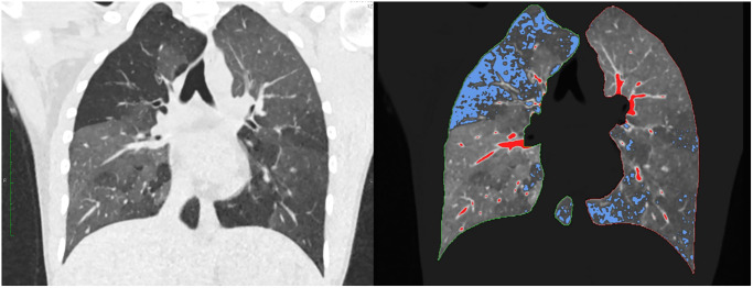
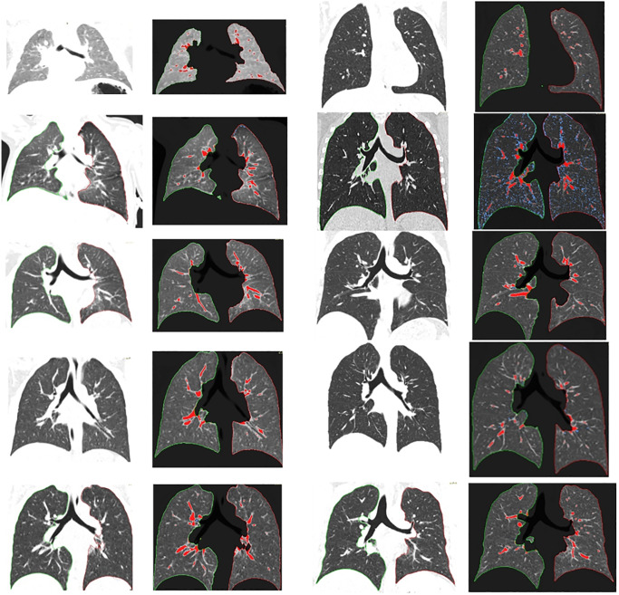
Dose-optimized chest CT examinations performed over the last 3 years using a dual-source CT were retrospectively analysed. Exclusion criteria were age >30 years and any known or newly diagnosed lung pathology. Lung volume, mean lung density, full-width-at-half-maximum and low attenuated volume (LAV) were semi-automated quantified in 151 patients. qCT values between different age groups as well as unenhanced (Group 1) and contrast-enhanced (Group 2) protocols were compared. Models for projection of age-dependant changes in qCT values were fitted.
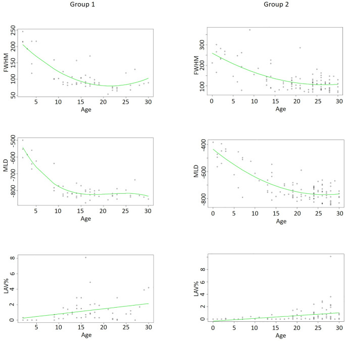
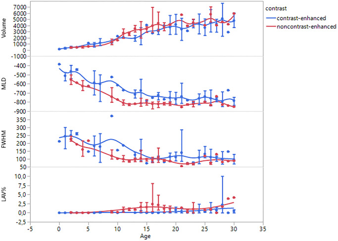
Significant differences in qCT parameters were found between the age groups from 0 to 15 years (p < 0.05). All parameters except LAV merge into a plateau level above this age as shown by polynomial models (r2 between 0.85 and 0.67). In group 2, this plateau phase is shifted back around five years. Except for the volume, significant differences in all qCT values were found between group 1 and 2 (p < 0.01).
qCT parameters underly a specific age-dependant dynamic. Except for LAV, qCT parameters reach a plateau around adolescence. Contrast-enhanced protocols seem to shift this plateau backwards.
定量计算机断层扫描(qCT)与气流阻塞和肺部纤维化改变相关。然而,由于 qCT 研究通常集中在患病和老年患者身上,目前的文献缺乏身体发育过程中的生理 qCT 值。我们评估了健康队列的胸部 CT 检查,从婴儿期到成年期,以确定生理 qCT 值和身体发育过程中的变化。
回顾性分析了过去 3 年使用双源 CT 进行的剂量优化胸部 CT 检查。排除标准为年龄>30 岁和任何已知或新诊断的肺部病理学。在 151 名患者中,使用半自动方法对肺容积、平均肺密度、半高全宽和低衰减体积(LAV)进行定量。比较了不同年龄组之间以及未增强(第 1 组)和增强(第 2 组)方案之间的 qCT 值。拟合了用于预测 qCT 值与年龄相关变化的模型。
在 0 至 15 岁的年龄组之间发现 qCT 参数存在显著差异(p <0.05)。除 LAV 外,所有参数在该年龄以上均呈平台水平,如多项式模型所示(r2 介于 0.85 和 0.67 之间)。在第 2 组中,该平台阶段向后移动了大约五年。除体积外,第 1 组和第 2 组之间的所有 qCT 值均存在显著差异(p <0.01)。
qCT 参数具有特定的年龄依赖性动态。除 LAV 外,qCT 参数在青春期左右达到平台。增强协议似乎会将此平台向后移动。