Experimental Rheumatology, Radboud university medical center, P.O. Box 9101, 6500 HB Nijmegen, the Netherlands.
Department of Oral Cell Biology and Functional Anatomy, Academic Centre for Dentistry Amsterdam (ACTA), University of Amsterdam and Vrije Universiteit Amsterdam, Gustav Mahlerlaan 3004, 1081 LA Amsterdam, the Netherlands.
Int J Mol Sci. 2020 May 27;21(11):3774. doi: 10.3390/ijms21113774.
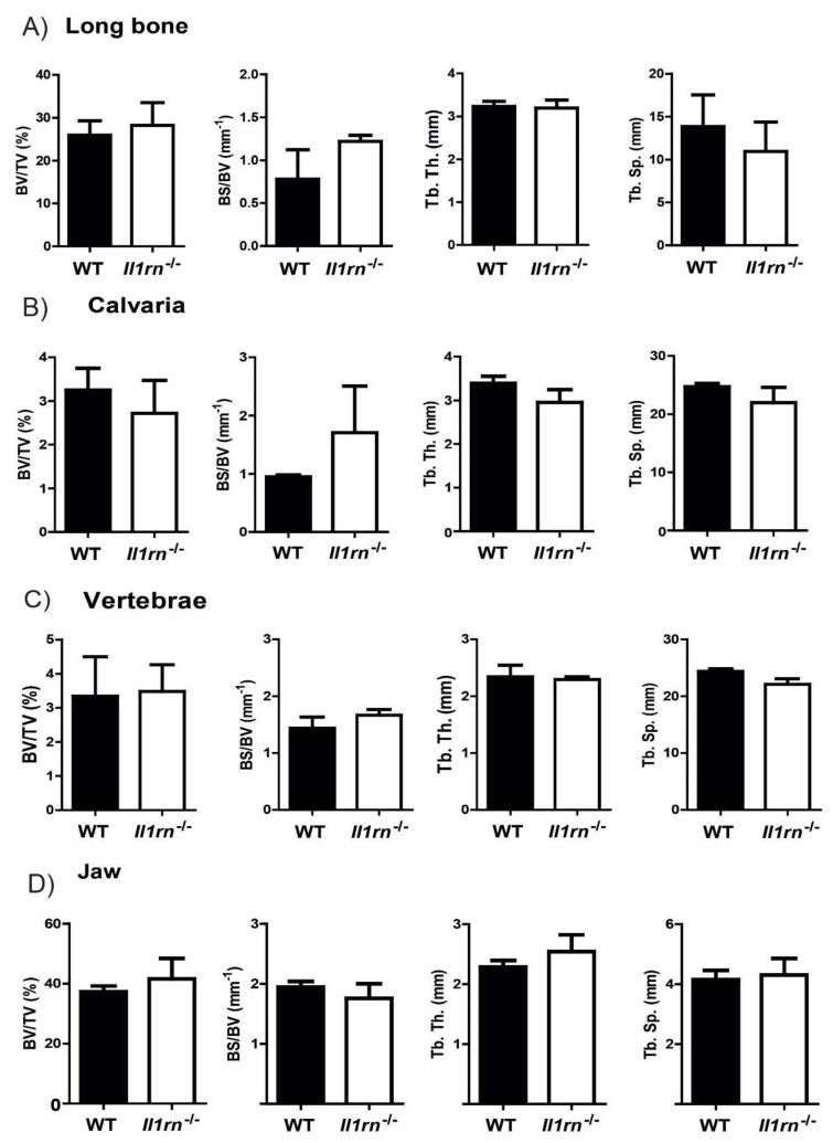
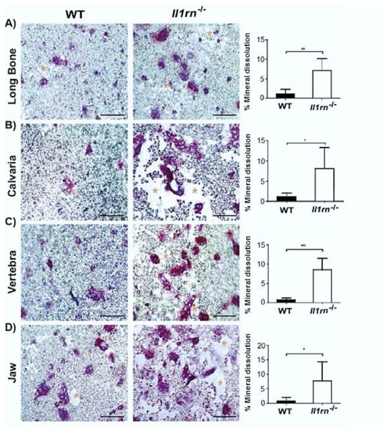
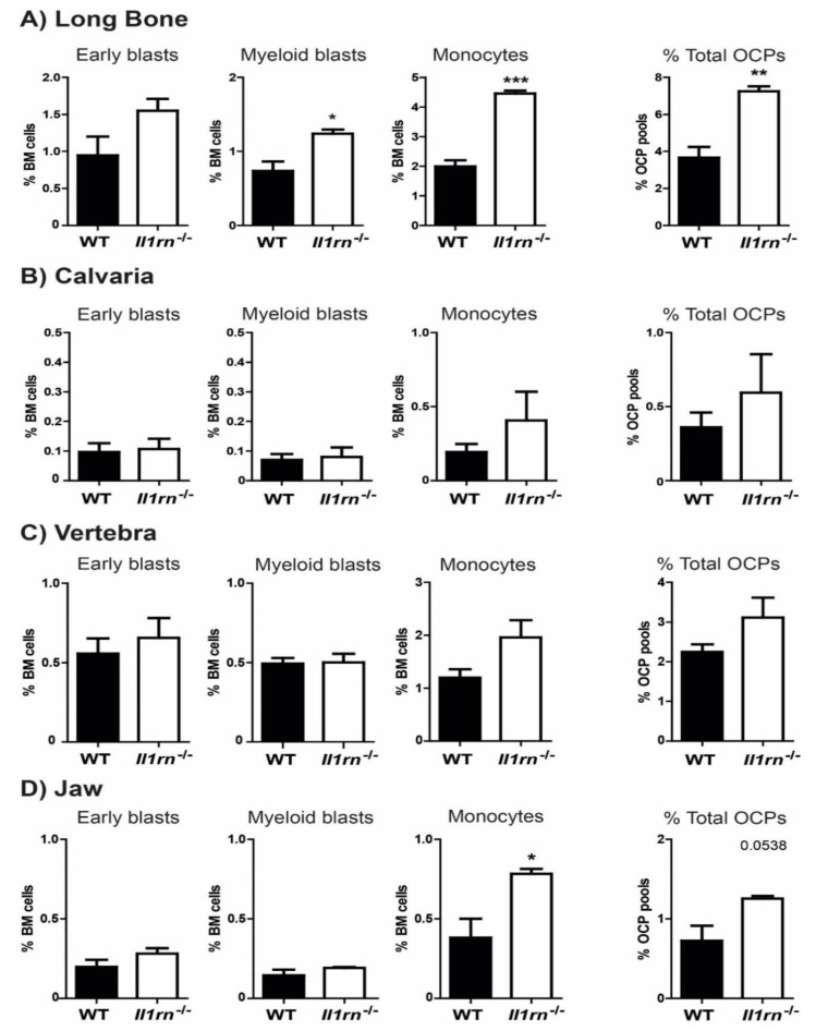
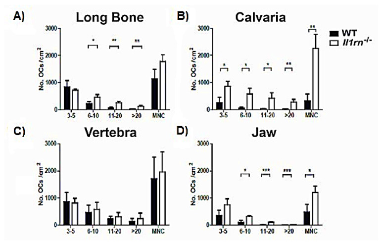
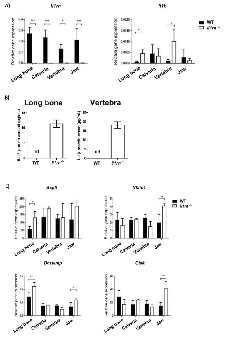
Recently, it was shown that interleukin-1β (IL-1β) has diverse stimulatory effects on different murine long bone marrow osteoclast precursors (OCPs) in vitro. In this study, interleukin-1 receptor antagonist deficient () and wild-type (WT) mice were compared to investigate the effects of enhanced IL-1 signaling on the composition of OCPs in long bone, calvaria, vertebra, and jaw. Bone marrow cells were isolated from these sites and the percentage of early blast (CD31 Ly-6C), myeloid blast (CD31 Ly-6C), and monocyte (CD31 Ly-6C) OCPs was assessed by flow cytometry. At the time-point of cell isolation, mice showed no inflammation or bone destruction yet as determined by histology and microcomputed tomography. However, mice had an approximately two-fold higher percentage of OCPs in long bone and jaw marrow compared to WT. Conversely, vertebrae and calvaria marrow contained a similar composition of OCPs in both strains. Bone marrow cells were cultured with macrophage colony stimulating factor (M-CSF) and receptor of NfκB ligand (RANKL) on bone slices to assess osteoclastogenesis and on calcium phosphate-coated plates to analyze mineral dissolution. Deletion of increased osteoclastogenesis from long bone, calvaria, and jaw marrows, and all cultures showed increased mineral dissolution compared to WT. However, osteoclast markers increased exclusively in osteoclasts from long bone and jaw. Collectively, these findings indicate that a lack of IL-1RA increases the numbers of OCPs in vivo, particularly in long bone and jaw, where rheumatoid arthritis and periodontitis develop. Thus, increased bone loss at these sites may be triggered by a larger pool of OCPs due to the disruption of IL-1 inhibitors.
最近,研究表明白细胞介素-1β(IL-1β)在体外对不同的鼠长骨髓破骨细胞前体(OCPs)具有多种刺激作用。在这项研究中,比较了白细胞介素-1受体拮抗剂缺陷()和野生型(WT)小鼠,以研究增强的 IL-1 信号对长骨、颅骨、脊柱和颌骨中 OCPs 组成的影响。从这些部位分离骨髓细胞,并通过流式细胞术评估早期 blast(CD31 Ly-6C)、髓样 blast(CD31 Ly-6C)和单核细胞(CD31 Ly-6C)OCPs 的百分比。在细胞分离时,尽管组织学和微计算机断层扫描未显示出炎症或骨破坏,但 小鼠的长骨和颌骨髓中 OCPs 的百分比约为 WT 的两倍。相反,两种菌株的椎骨和颅骨骨髓中 OCPs 的组成相似。用巨噬细胞集落刺激因子(M-CSF)和核因子 kappa B 配体受体(RANKL)在骨片上培养骨髓细胞以评估破骨细胞生成,并在磷酸钙涂层板上分析矿物质溶解。 的缺失增加了长骨、颅骨和颌骨骨髓中的破骨细胞生成,所有 培养物的矿物质溶解均比 WT 增加。然而,破骨细胞标志物仅在长骨和颌骨的 破骨细胞中增加。总之,这些发现表明,IL-1RA 的缺乏增加了体内 OCPs 的数量,特别是在类风湿关节炎和牙周炎发生的长骨和颌骨中。因此,由于 IL-1 抑制剂的破坏,这些部位的骨丢失增加可能是由于 OCPs 池的增加所触发的。