Tanaka Koji, Matsumoto Shoji, Yamada Takeshi, Yamasaki Ryo, Suzuki Makoto, Kido Mizuho A, Kira Jun-Ichi
Department of Neurology, Neurological Institute, Graduate School of Medical Sciences, Kyushu University, Fukuoka, Japan.
Department of Comprehensive Strokology, School of Medicine, Fujita Health University, Toyoake, Japan.
Front Neurosci. 2020 May 12;14:453. doi: 10.3389/fnins.2020.00453. eCollection 2020.
In the acute phase of ischemia-reperfusion, hypoperfusion associated with ischemia and reperfusion in microvascular regions and disruption of the blood-brain barrier (BBB) contribute to post-ischemic brain injury. We aimed to clarify whether brain injury following transient middle cerebral artery occlusion (tMCAO) is ameliorated in knockout ( ) mice.
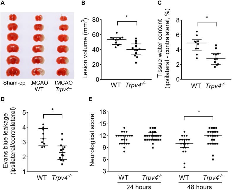
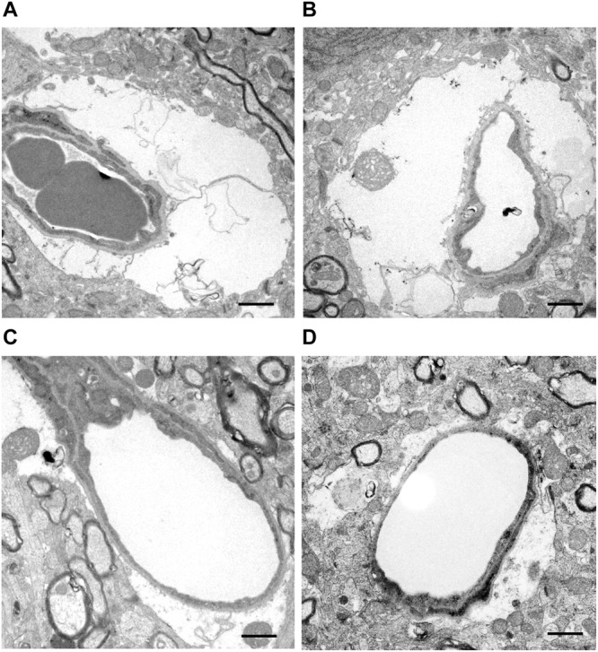
tMCAO was induced in wild-type (WT) and mice aged 8-10 weeks. Ischemia-induced lesion volume was evaluated by 2,3,5-triphenyltetrazolium chloride staining at 24 h post-tMCAO. Tissue water content and Evans blue leakage in the ipsilateral hemisphere and a neurological score were evaluated at 48 h post-tMCAO. Transmission electron microscopy (TEM) was performed to assess the morphological changes in microvasculature in the ischemic lesions at 6 h post-tMCAO.
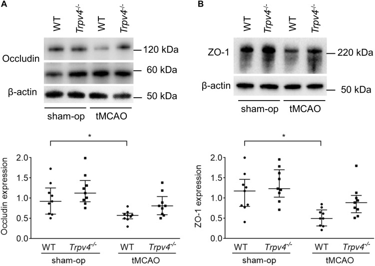
Compared with WT mice, mice showed reduced ischemia-induced lesion volume and reduced water content and Evans blue leakage in the ipsilateral hemisphere alongside milder neurological symptoms. The loss of zonula occludens-1 and occludin proteins in the ipsilateral hemisphere was attenuated in mice. TEM revealed that parenchymal microvessels in the ischemic lesion were compressed and narrowed by the swollen endfeet of astrocytes in WT mice, but these effects were markedly ameliorated in mice.
The present results demonstrate that TRPV4 contributes to post-ischemic brain injury. The preserved microcirculation and BBB function shortly after reperfusion are the key neuroprotective roles of TRPV4 inhibition, which represents a promising target for the treatment of acute ischemic stroke.
在缺血再灌注的急性期,微血管区域与缺血和再灌注相关的低灌注以及血脑屏障(BBB)的破坏会导致缺血后脑损伤。我们旨在阐明在敲除( )小鼠中短暂性大脑中动脉闭塞(tMCAO)后脑损伤是否得到改善。
对8 - 10周龄的野生型(WT)和 小鼠进行tMCAO诱导。在tMCAO后24小时通过氯化三苯基四氮唑染色评估缺血诱导的损伤体积。在tMCAO后48小时评估同侧半球的组织含水量、伊文思蓝渗漏情况以及神经学评分。在tMCAO后6小时进行透射电子显微镜(TEM)检查,以评估缺血性病变中微血管的形态变化。
与WT小鼠相比, 小鼠的缺血诱导损伤体积减小,同侧半球的含水量和伊文思蓝渗漏减少,神经症状较轻。 小鼠同侧半球中紧密连接蛋白-1和闭合蛋白的缺失得到减轻。TEM显示,WT小鼠缺血性病变中的实质微血管被星形胶质细胞肿胀的终足挤压和变窄,但在 小鼠中这些影响明显改善。
目前的结果表明TRPV4促成缺血后脑损伤。再灌注后不久保留的微循环和血脑屏障功能是TRPV4抑制的关键神经保护作用,这代表了急性缺血性中风治疗的一个有前景的靶点。