• 文献检索
  • 文档翻译
  • 深度研究
  • 学术资讯
  • Suppr Zotero 插件Zotero 插件
  • 邀请有礼
  • 套餐&价格
  • 历史记录
应用&插件
Suppr Zotero 插件Zotero 插件浏览器插件Mac 客户端Windows 客户端微信小程序
定价
高级版会员购买积分包购买API积分包
服务
文献检索文档翻译深度研究API 文档MCP 服务
关于我们
关于 Suppr公司介绍联系我们用户协议隐私条款
关注我们

Suppr 超能文献

核心技术专利:CN118964589B侵权必究
粤ICP备2023148730 号-1Suppr @ 2026

文献检索

告别复杂PubMed语法,用中文像聊天一样搜索,搜遍4000万医学文献。AI智能推荐,让科研检索更轻松。

立即免费搜索

文件翻译

保留排版,准确专业,支持PDF/Word/PPT等文件格式,支持 12+语言互译。

免费翻译文档

深度研究

AI帮你快速写综述,25分钟生成高质量综述,智能提取关键信息,辅助科研写作。

立即免费体验

化学诱导的人肝祖细胞来源的外泌体抑制氧化应激诱导的细胞死亡。

Exosomes derived from chemically induced human hepatic progenitors inhibit oxidative stress induced cell death.

机构信息

Center for Exosome & Bioparticulate Research, Hanyang University, Gyeonggi-do, Korea.

HY Indang Center of Regenerative Medicine and Stem Cell Research, Hanyang University, Seoul, Korea.

出版信息

Biotechnol Bioeng. 2020 Sep;117(9):2658-2667. doi: 10.1002/bit.27447. Epub 2020 Jun 30.

DOI:10.1002/bit.27447
PMID:32484909
原文链接:https://pmc.ncbi.nlm.nih.gov/articles/PMC7496643/
Abstract

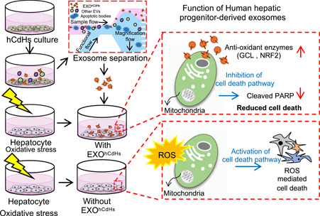
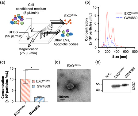
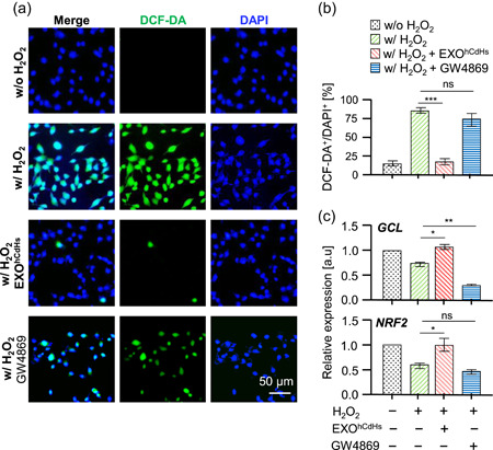
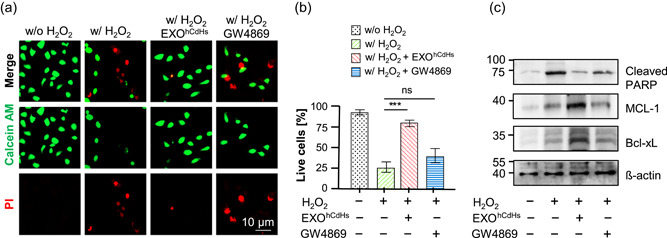
The emerging field of regenerative medicine has revealed that the exosome contributes to many aspects of development and disease through intercellular communication between donor and recipient cells. However, the biological functions of exosomes secreted from cells have remained largely unexplored. Here, we report that the human hepatic progenitor cells (CdHs)-derived exosome (EXO ) plays a crucial role in maintaining cell viability. The inhibition of exosome secretion treatment with GW4869 results in the acceleration of reactive oxygen species (ROS) production, thereby causing a decrease of cell viability. This event provokes inhibition of caspase dependent cell death signaling, leading to a ROS-dependent cell damage response and thus induces promotion of antioxidant gene expression or repair of cell death of hypoxia-exposed cells. Together, these findings show the effect of exosomes in regeneration of liver cells, and offer valuable new insights into liver regeneration.

摘要

再生医学领域的新发现表明,外泌体通过供体细胞与受体细胞之间的细胞间通讯,在许多方面发挥作用,参与到发育和疾病过程中。然而,细胞分泌的外泌体的生物学功能在很大程度上仍未得到探索。在这里,我们报告称,人肝祖细胞(CdHs)来源的外泌体(EXO)在维持细胞活力方面起着关键作用。用 GW4869 抑制外泌体分泌处理会加速活性氧(ROS)的产生,从而导致细胞活力下降。这一事件引发了半胱天冬酶依赖性细胞死亡信号的抑制,导致 ROS 依赖性细胞损伤反应,从而诱导抗氧化基因表达的促进或缺氧暴露细胞死亡的修复。总之,这些发现表明了外泌体在肝细胞再生中的作用,并为肝脏再生提供了有价值的新见解。

https://cdn.ncbi.nlm.nih.gov/pmc/blobs/6d27/7496643/b58fcb69ae1c/BIT-117-2658-g005.jpg
https://cdn.ncbi.nlm.nih.gov/pmc/blobs/6d27/7496643/4e53cce40d61/BIT-117-2658-g001.jpg
https://cdn.ncbi.nlm.nih.gov/pmc/blobs/6d27/7496643/30c4a720ba5d/BIT-117-2658-g002.jpg
https://cdn.ncbi.nlm.nih.gov/pmc/blobs/6d27/7496643/28dc50c0f4e7/BIT-117-2658-g003.jpg
https://cdn.ncbi.nlm.nih.gov/pmc/blobs/6d27/7496643/73e6d1a7b135/BIT-117-2658-g004.jpg
https://cdn.ncbi.nlm.nih.gov/pmc/blobs/6d27/7496643/b58fcb69ae1c/BIT-117-2658-g005.jpg

相似文献

1
Exosomes derived from chemically induced human hepatic progenitors inhibit oxidative stress induced cell death.化学诱导的人肝祖细胞来源的外泌体抑制氧化应激诱导的细胞死亡。
Biotechnol Bioeng. 2020 Sep;117(9):2658-2667. doi: 10.1002/bit.27447. Epub 2020 Jun 30.
2
Small molecule-mediated reprogramming of human hepatocytes into bipotent progenitor cells.小分子介导的人肝细胞重编程为双能祖细胞。
J Hepatol. 2019 Jan;70(1):97-107. doi: 10.1016/j.jhep.2018.09.007. Epub 2018 Sep 19.
3
Exosomes maintain cellular homeostasis by excreting harmful DNA from cells.外泌体通过排出细胞内有害的 DNA 来维持细胞内环境的稳定。
Nat Commun. 2017 May 16;8:15287. doi: 10.1038/ncomms15287.
4
Palladium Nanoparticle-Induced Oxidative Stress, Endoplasmic Reticulum Stress, Apoptosis, and Immunomodulation Enhance the Biogenesis and Release of Exosome in Human Leukemia Monocytic Cells (THP-1).钯纳米颗粒诱导的氧化应激、内质网应激、细胞凋亡和免疫调节增强人单核白血病细胞(THP-1)中外泌体的生物发生和释放。
Int J Nanomedicine. 2021 Apr 15;16:2849-2877. doi: 10.2147/IJN.S305269. eCollection 2021.
5
Exosomes from Human-Induced Pluripotent Stem Cell-Derived Mesenchymal Stromal Cells (hiPSC-MSCs) Protect Liver against Hepatic Ischemia/ Reperfusion Injury via Activating Sphingosine Kinase and Sphingosine-1-Phosphate Signaling Pathway.人诱导多能干细胞来源的间充质基质细胞(hiPSC-MSCs)分泌的外泌体通过激活鞘氨醇激酶和1-磷酸鞘氨醇信号通路保护肝脏免受肝缺血/再灌注损伤。
Cell Physiol Biochem. 2017;43(2):611-625. doi: 10.1159/000480533. Epub 2017 Sep 21.
6
Exosome-Mediated Intercellular Communication between Hepatitis C Virus-Infected Hepatocytes and Hepatic Stellate Cells.丙型肝炎病毒感染的肝细胞与肝星状细胞之间通过外泌体介导的细胞间通讯
J Virol. 2017 Feb 28;91(6). doi: 10.1128/JVI.02225-16. Print 2017 Mar 15.
7
miR-122 Release in Exosomes Precedes Overt Tolvaptan-Induced Necrosis in a Primary Human Hepatocyte Micropatterned Coculture Model.外泌体中 miR-122 的释放先于原发性人肝细胞微图案共培养模型中托伐普坦诱导的坏死。
Toxicol Sci. 2018 Jan 1;161(1):149-158. doi: 10.1093/toxsci/kfx206.
8
Delphinidin protects β2m-/Thy1+ bone marrow-derived hepatocyte stem cells against TGF-β1-induced oxidative stress and apoptosis through the PI3K/Akt pathway in vitro.飞燕草素通过 PI3K/Akt 通路体外保护β2m-/Thy1+骨髓源性肝干细胞免受 TGF-β1 诱导的氧化应激和细胞凋亡。
Chem Biol Interact. 2019 Jan 5;297:109-118. doi: 10.1016/j.cbi.2018.10.019. Epub 2018 Oct 23.
9
Cardiac progenitor cell-derived exosomes prevent cardiomyocytes apoptosis through exosomal miR-21 by targeting PDCD4.心脏祖细胞衍生的外泌体通过靶向PDCD4的外泌体miR-21预防心肌细胞凋亡。
Cell Death Dis. 2016 Jun 23;7(6):e2277. doi: 10.1038/cddis.2016.181.
10
The endocannabinoid N-arachidonoyl dopamine (NADA) selectively induces oxidative stress-mediated cell death in hepatic stellate cells but not in hepatocytes.内源性大麻素 N-花生四烯酰多巴胺(NADA)选择性诱导肝星状细胞发生氧化应激介导的细胞死亡,而不诱导肝细胞发生这种死亡。
Am J Physiol Gastrointest Liver Physiol. 2012 Apr 15;302(8):G873-87. doi: 10.1152/ajpgi.00241.2011. Epub 2012 Feb 2.

引用本文的文献

1
Exosomes from liver progenitor cells carrying JAG1 activate notch signaling to promote liver regeneration in PVL rats.携带JAG1的肝祖细胞来源的外泌体激活Notch信号通路以促进门静脉结扎肝切除大鼠的肝再生。
Cell Death Dis. 2025 Aug 12;16(1):609. doi: 10.1038/s41419-025-07925-1.
2
Characteristics of chemically induced liver progenitors derived from a pig model of metabolic dysfunction-associated steatotic liver disease.源自代谢功能障碍相关脂肪性肝病猪模型的化学诱导肝祖细胞的特征
PLoS One. 2024 Dec 5;19(12):e0313312. doi: 10.1371/journal.pone.0313312. eCollection 2024.
3
Engineering extracellular vesicles for ROS scavenging and tissue regeneration.

本文引用的文献

1
Understanding the marvels behind liver regeneration.了解肝脏再生背后的奇迹。
Wiley Interdiscip Rev Dev Biol. 2019 May;8(3):e340. doi: 10.1002/wdev.340. Epub 2019 Mar 28.
2
Exosomes: biogenesis, biologic function and clinical potential.外泌体:生物发生、生物学功能及临床应用潜力
Cell Biosci. 2019 Feb 15;9:19. doi: 10.1186/s13578-019-0282-2. eCollection 2019.
3
Augmenter of liver regeneration: Essential for growth and beyond.肝再生增强因子:生长所必需,不止于此。
工程化细胞外囊泡用于活性氧清除和组织再生。
Nano Converg. 2024 Jun 26;11(1):24. doi: 10.1186/s40580-024-00430-9.
4
Autophagy in hepatic progenitor cells modulates exosomal miRNAs to inhibit liver fibrosis in schistosomiasis.肝祖细胞中的自噬通过调节外泌体 miRNAs 抑制血吸虫病肝纤维化。
Front Med. 2024 Jun;18(3):538-557. doi: 10.1007/s11684-024-1079-1. Epub 2024 May 21.
5
Antioxidant Iron Oxide Nanoparticles: Their Biocompatibility and Bioactive Properties.抗氧化铁氧化物纳米粒子:它们的生物相容性和生物活性特性。
Int J Mol Sci. 2023 Nov 2;24(21):15901. doi: 10.3390/ijms242115901.
6
Emerging role of engineered exosomes in nonalcoholic fatty liver disease.工程化外泌体在非酒精性脂肪性肝病中的新兴作用。
World J Hepatol. 2023 Mar 27;15(3):386-392. doi: 10.4254/wjh.v15.i3.386.
7
Transplantation of chemically-induced liver progenitor cells ameliorates hepatic fibrosis in mice with diet-induced nonalcoholic steatohepatitis.化学诱导的肝祖细胞移植可改善饮食诱导的非酒精性脂肪性肝炎小鼠的肝纤维化。
Regen Ther. 2022 Nov 25;21:574-583. doi: 10.1016/j.reth.2022.11.001. eCollection 2022 Dec.
8
Crosstalk between Oxidative Stress and Exosomes.氧化应激与外泌体的串扰。
Oxid Med Cell Longev. 2022 Aug 30;2022:3553617. doi: 10.1155/2022/3553617. eCollection 2022.
9
Emerging Antioxidant Paradigm of Mesenchymal Stem Cell-Derived Exosome Therapy.新兴的间充质干细胞衍生外泌体治疗的抗氧化策略。
Front Endocrinol (Lausanne). 2021 Nov 29;12:727272. doi: 10.3389/fendo.2021.727272. eCollection 2021.
10
Secretome of Stem Cells: Roles of Extracellular Vesicles in Diseases, Stemness, Differentiation, and Reprogramming.干细胞的胞外囊泡组:细胞外囊泡在疾病、干性、分化和重编程中的作用。
Tissue Eng Regen Med. 2022 Feb;19(1):19-33. doi: 10.1007/s13770-021-00406-4. Epub 2021 Nov 24.
Cytokine Growth Factor Rev. 2019 Feb;45:65-80. doi: 10.1016/j.cytogfr.2018.12.003. Epub 2018 Dec 16.
4
Hepatic progenitors in liver regeneration.肝脏再生中的肝祖细胞。
J Hepatol. 2018 Dec;69(6):1394-1395. doi: 10.1016/j.jhep.2018.03.004. Epub 2018 Nov 1.
5
Engineering mesenchymal stem cells to improve their exosome efficacy and yield for cell-free therapy.工程化间充质干细胞以提高其外泌体的疗效和产量用于无细胞治疗。
J Extracell Vesicles. 2018 Sep 26;7(1):1522236. doi: 10.1080/20013078.2018.1522236. eCollection 2018.
6
Small molecule-mediated reprogramming of human hepatocytes into bipotent progenitor cells.小分子介导的人肝细胞重编程为双能祖细胞。
J Hepatol. 2019 Jan;70(1):97-107. doi: 10.1016/j.jhep.2018.09.007. Epub 2018 Sep 19.
7
Analysis of nano-crystals: Evaluation of heavy metal-embedded biological specimen by high voltage electron microscopy.纳米晶体分析:通过高压电子显微镜对重金属嵌入生物样本进行评估。
Ultramicroscopy. 2018 Nov;194:35-39. doi: 10.1016/j.ultramic.2018.07.003. Epub 2018 Jul 10.
8
Exosome-Mimetic Nanovesicles from Hepatocytes promote hepatocyte proliferation in vitro and liver regeneration in vivo.肝细胞来源的外泌体模拟纳米囊泡促进体外肝细胞增殖和体内肝再生。
Sci Rep. 2018 Feb 6;8(1):2471. doi: 10.1038/s41598-018-20505-y.
9
Liver Bioengineering Using Decellularized Whole-Liver Scaffolds.使用去细胞全肝支架的肝脏生物工程
Methods Mol Biol. 2018;1577:293-305. doi: 10.1007/7651_2017_98.
10
Separation of extracellular nanovesicles and apoptotic bodies from cancer cell culture broth using tunable microfluidic systems.采用可调谐微流控系统从癌细胞培养液中分离细胞外纳米囊泡和凋亡小体。
Sci Rep. 2017 Aug 30;7(1):9907. doi: 10.1038/s41598-017-08826-w.